


##issue.toc##
##article.goto##
https://doi.org/10.21886/2219-8075-2021-12-2-48-53
##article.goto##
Objective: to conduct a comparative analysis of the structure, number, and antenatal diagnosis of fetal malformations in two clinical groups. Materials and methods: a retrospective comparative analysis of the variations, frequency, and detectability of malformations and developmental abnormalities in newborns born in two groups was conducted for the periods from 2010 to 2012 (the first group) and from 2017 to 2019 (the second group). The study of these materials was conducted on the basis of the maternity department of the city hospital of Rostov-on-Don. Statistical processing of the obtained results was carried out using the programs Statistica 10 and Microsoft Excel 2013. Results: in the second group, the number of children with genital abnormalities increased. The number of children with skin abnormalities increased as well as the number of children with gastrointestinal malformations. There is a decrease in the number of abnormalities in the development of the musculoskeletal system and the circulatory system. The rate of cleavage of the lip and hard palate decreased. However, malformations of the fetus were not detected in all cases, although their detectability increased from 4.41% in 2010 – 2012 to 22.62% in 2017 – 2019, i.e. by 5.13 times. Conclusion. The search for modern, reliable, and non-invasive methods for diagnosing fetal malformations in early pregnancy is an important stage in the study of this issue. It is also important to train high-class ultrasound specialists as well as search for new biochemical markers of congenital genetic disorders.
Pertseva G.M., Borscheva A.A., Alekseeva N.A. Comparative analysis of congenital malformations of the fetus detected in the periods from 2010 to 2012 and from 2017 to 2019 (based on the materials of the maternity department of Rostov-on-Don). Medical Herald of the South of Russia. 2021;12(2):48-53. (In Russ.) https://doi.org/10.21886/2219-8075-2021-12-2-48-53
Every year, congenital malformations in children are leading among both medical and social problems of the modern world [1, 2]. Even in ancient times, scientists were interested in the reasons for the birth of children with various types of birth defects [2]. In Russia, the beginning of teratology development is considered to be the year 1718, when the teratomorph museum was created according to Peter the Great’s decree [3, 4]. However, the intensive development of Russian teratology started in the 1980s. Fetal malformations are one of the most threatening complications that lead to the fetus and newborn disability and mortality. They took the 2nd-3rd places in the structure of causes of the perinatal fetus and newborn death [2]. The factors that provoke the development of malformations in the fetus and newborn are diverse. A combination of social, economic, medical, and genetic causes can be a trigger for the occurrence of fetal malformations [5]. However, a number of authors [6] believe that the exact causes of abnormal fetal development in 80% of cases remain unknown. Only 10–15% of deformities may be explained by a genetic factor, at a time when drug aggression accounts for 1–5%. As for the development of any malformation type, it is important to note that the key point is the stage of intrauterine fetus development, but not the impact factor itself. The intensity of pathological fetus development depends on the location of the genetic breakdown and the strength of the toxic effect. However, there is an opinion that there is no clear correlation between the strength of the negative factor and the intensity of malformations [2, 5, 6]. There are often cases of children born with severe malformations in young, healthy parents with a favorable genetic history, who did not have bad habits. There are certain criteria, on the basis of which various classifications of fetal malformations are compiled; they differ from each other in the time of the negative factor influence. These are gametopathies, blastopathies, embryopathies, and fetopathies. Another classification of fetal malformations reflects their types, such as agnosia, hypoplasia, heterotopia, ectopia, chromosomal diseases, etc. There is also a classification, according to which malformations are divided into surgically cured ones, compatible with life, but provoking serious problems, and malformations that are not compatible with life. One of the important aspects in teratology development is the prenatal diagnosis of various fetal malformations [5, 7]. After all, the birth of a child with obvious and especially uncorrectable malformations is a terrible tragedy for relatives and friends. Unfortunately, modern methods of medical examination do not always allow specialists to determine the presence of fetal malformations in the early gestational age. Therefore, the search for non-invasive, reliable methods for diagnosing fetal malformations in early pregnancy is the main direction in this complex problem. The early detection of fetal development abnormalities would allow finding the timely resolution concerning the issue of whether it is expedient to prolong pregnancy [3]. In this regard, such a problem is relevant, especially for obstetricians and gynecologists, neonatologists, and geneticists.
The purpose of the study is to conduct a comparative analysis of the structure, number, and antenatal diagnosis of fetal malformations in two clinical groups during the period from 2010 to 2012 and from 2017 to 2019.
A comparative analysis was carried out. It allowed studying the number and detectability of various fetal malformation types in women who gave birth (the maternity department of the Rostov-on-Don City Hospital) to children with abnormalities of the structure of certain organs during the periods of 2010–2012 (first group, 204 children) and 2017–2019 (second group, 168 children). The analysis was carried out according to the data of ultrasound protocols, screening genetic studies conducted for women of certain gestational age, also in accordance with the Order of the Ministry of Health of the Russian Federation dated November 01, 2012 No. 572n “On approval of the procedure for providing medical care in the field of “obstetrics and gynecology” (except for the use of assisted reproductive technologies)”. Also, the authors of the article used materials taken from the birth histories and development histories of children born. The ultrasound and Doppler-metric studies of the pelvic organs with the determination of fetal weight and size, placenta location, placenta structure, amniotic fluid index, and other mandatory parameters were conducted using the APLIO MX ultrasound diagnostic system (manufactured by TOSHIBA MEDICAL SISTEM, Japan, 2015) and using transvaginal and abdominal sensors, with a frequency of 5 and 6.5 MHz in grayscale modes. The cardiocotography study in childbirth was conducted with G6B Plus medical mother and fetus monitors (2017). Each exchange card and birth history had an informed consent signed by the woman to the processing of personal data. The statistical processing of the obtained results was carried out by means of the programs Statistica 10 and Microsoft Excel 2013. Therefore, the prevalence of signs (in %) was calculated. The statistical significance of differences in the prevalence of signs was evaluated by the Pearson chi-squared test. The differences between the groups were considered significant at a significance level of p < 0.05.
Thus, 11,048 children were born in the period from 2010 to 2012, of which 204 (1.85%) had developmental abnormalities. From 2017 to 2019, 168 (1.43%) children (p = 0.0134) of 11,716 born were found to have developmental defects (Table 1).
Table 1
Birth of children with defects in 2010–2012 and 2017–2019
|
Groups |
First group 2010–2012 |
Second group 2017–2019 |
|
Total number of births |
11,048 |
11,716 |
|
The number of children born with malformations during these periods |
204 – 1.85% |
168 – 1.43% |
While comparing the varieties of fetal malformations in the two study groups, it was noted that the malformations types were identical (Fig. 1).
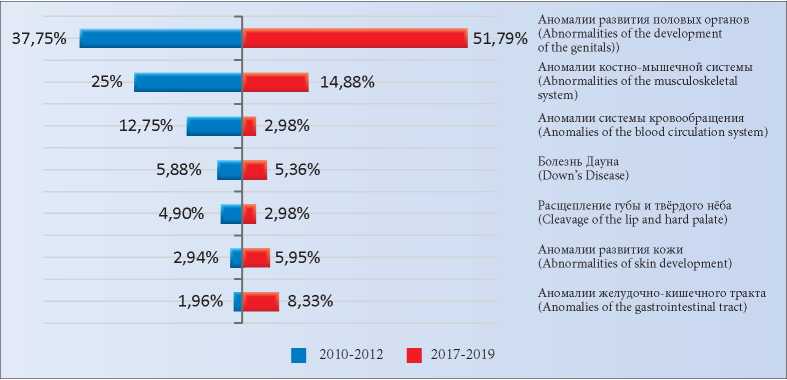
Fig. 1. Types of fetal malformations for 2010–2012 (first group) and 2017–2019 (second group).
The percentage is calculated from the total number of all the detected anomalies.
Therefore, interesting facts were revealed while comparing the malformations types in fetuses of the two clinical groups. Therefore, both in 2010–2012 and in 2017–2019, malformations of the genitals and musculoskeletal system predominated. However, over the past three years, the number of children with genital abnormalities has increased. If in the period from 2010 to 2012 there were 77 (37.75%) cases, then in 2017–2019, 87 (51.79%) children were born with such an anomaly (p = 0.00664). The number of children with skin abnormalities increased slightly – from 6 (2.94%) to 10 (5.95%) (p = 0.15427). The number of children with gastrointestinal tract defects increased from 4 (1.96%) to 14 (8.33%) (p = 0.004365). There was also a decrease in the number of musculoskeletal system abnormalities – from 51 (25.00%) to 25 (14.88%) (p = 0.015998), the circulatory system abnormalities – from 26 (12.75%) to 5 (2.98%) (p = 0.000692). Such pathology as cleavage of the lip and hard palate decreased from 10 (4.90%) to 5 (2.98%) (p = 0.347397). The number of children with Down's disease decreased slightly. In 2010–2012, there were 12 (5.88%), and in 2017–2019 – 9 (5.36%) (p = 0.827095). All these data are presented in Table 2.
Table 2
Quantitative equivalent of detected fetal malformations for 2010–2012 and 2017–2019
|
Types of fetal malformations |
2010–2012 First group |
2017–2019 Second group |
Significance level of differences (p) |
|
Abnormalities of the development of the genitals |
77 (37.75%) |
87 (51.79%) |
0.00664
|
|
Abnormalities of skin development |
6 (2.94%) |
10 (5.95%) |
015427
|
|
Anomalies of the gastrointestinal tract |
4 (1.96%) |
14 (8.33%) |
0004365
|
|
Abnormalities of the musculoskeletal system |
51(25%) |
25 (14.88%) |
0015998
|
|
Anomalies of the blood circulation system |
26 (12.75%) |
5 (2.98%) |
0.000692
|
|
Cleavage of the lip and hard palate |
10 (4.9%) |
5 (2.98%) |
0.347397
|
|
Down's disease |
12 (5.88%) |
9 (5.36%) |
0.827095
|
All the pregnant women of both clinical groups starting from the early gestational age were constantly supervised by doctors of the maternity welfare clinic and were examined in accordance with the Order of the Ministry of Health of the Russian Federation dated November 1, 2012. No. 572n “On approval of the procedure for providing medical care in the field of “obstetrics and gynecology” (except for the use of assisted reproductive technologies)”. However, fetal malformations were not detected in all the cases. Thus, only 9 (4.41%) of 204 children born in the first clinical group were diagnosed as ones with malformations during pregnancy, and 195 (95.59%) newborns were diagnosed with developmental abnormalities after birth. In the second clinical group, 38 (22.62%) of 168 children born had malformations detected intra-uterine, and 130 (77.38%) had malformations detected only after their birth (Figs. 2 and 3) (p = 0.0000001).
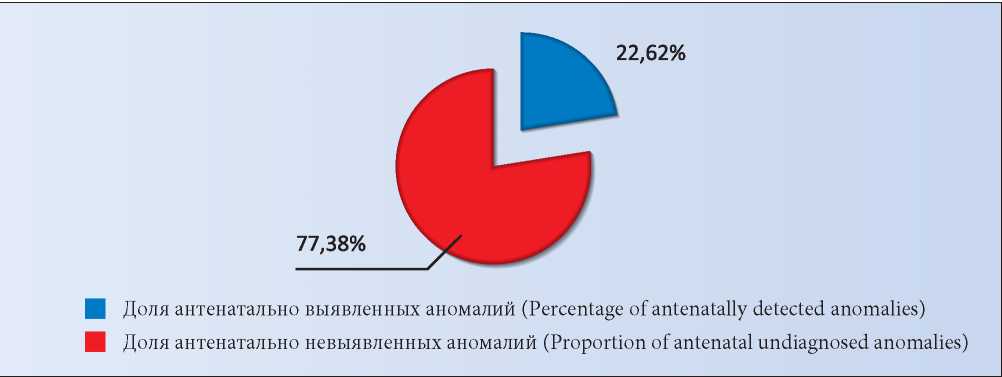
Fig. 2. Detection of intravenous fetal malformations (first group) in 2010–2012.
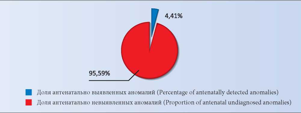
Fig. 3. Detection of intravenous fetal malformations (second group) in 2017–2019.
As it may be seen in Figs. 2 and 3, the detection of malformations for the period of 2010–2012 (in the first group) was 4.41%, and for the period of 2017–2019 (in the second group) was 22.62% (p = 0.0000001). However, despite the fact that the detection of malformations in 2017–2019 increased by 5.13 times, it still remains a very low one.
The obtained data suggest that heart defects, abnormalities in the musculoskeletal system development, and malformations of the gastrointestinal tract were mainly detected. Other fetal developmental defects were rarely detected intra-uterine and were only detected after childbirth. It should also be noted that the prenatal diagnosis of fetal abnormalities was more informative at the second medical examination screening of pregnant women (at 20–22 weeks). The prenatal diagnosis of heart defects and gastrointestinal tract abnormalities allowed performing the surgical correction immediately after the childbirth, which made it possible for these children to survive with subsequent satisfactory development.
The priority task of the state is the protection of motherhood and childhood. As can be seen from the data mentioned above, the percentage of children born in these periods with various types of defects is consistently in the range of 1.45–1.85%, and their detection in the prenatal period, although increasing, still remains very low. A comprehensive medical examination of pregnant women and constant monitoring in the maternity welfare clinics, unfortunately, contributes to a small increase in the prenatal diagnosis of fetal malformations. Unfortunately, even the revealed defects were diagnosed at a late date, that is, about after 20 weeks of pregnancy.
The structure of congenital malformations did not change during the mentioned above periods. An increase in the number of malformations of the reproductive system, skin, and gastrointestinal tract was noted as well. The number of malformations of the musculoskeletal and cardiovascular systems decreased. The percentage of children with Down syndrome remains stable. It should also be noted that despite the increase in the malformations detection in the antenatal period by 5.13 times, its percentage remains low. Thus, this problem may be considered as a still relevant one, especially the aspects of high-quality prenatal diagnostics are relevant nowadays. It is important to note that even timely screening, including ultrasound and biochemical blood tests for the corresponding hormones, does not always lead to a successful result. The detection of anomalies remains at a low level. The search for modern, reliable, non-invasive methods for diagnosing fetal malformations in early pregnancy is an important stage in the study of this complex and multifaceted problem. Also, it is urgent to train and teach high-class ultrasound specialists, as well as to search for new biochemical markers of congenital genetic breakdowns. Timely and comprehensive medical examination of pregnant women and the use of new highly informative research methods should lead to an increase in the percentage of fetal abnormalities detectability. This problem is especially relevant in the risk groups that should be formed in the process of pre-conceptional examination and women's preparation for planned pregnancy.
1. Alferovich E.N., Grak L.V. Embryophetopathy in newborns. Study guide. Ministry of Health of the Republic of Belarus. Minsk BSMU; 2017. (In Russ).
2. Voevodina S.M., Shemanaeva T.V. Prevention of congenital malformations in the fetus (literature review). Current problems of health care and medical statistics. 2018;(2):86-93. (In Russ). eLIBRARY ID: 35191961
3. Nagorneva S.V., Prokhorova V.S., Shelaeva E.V., Khudovecova A.M. The prevalence of congenital fetal anomalies for the past 5 years (2013–2017). Journal of Obstetrics and Women’s Diseases. 2018;67(3):44-48. (In Russ.). DOI: 10.17816/JOWD67344-48
4. Alekseeva R.L. Congenital malformations, prevention. Forum of Medical Sciences Tyumen State Medical University. 2017:48-51. (In Russ)
5. Novikova S.V., Zhuchenko L.A. Primary prevention of congenital malformations. Breast cancer. Mother and Child. 2015;23(1):25-28. (In Russ). eLIBRARY ID: 23772996
6. Pertceva G.M., Borscheva A.A., Kudinova E.E. Course of pregnancy and childbirth of a woman with achondroplasia. Kuban Scientific Medical Bulletin. 2017;1(2):163-165. (In Russ.). DOI: 10.25207/1608-6228-2017-2-163-165
7. Borscheva A.A., Pertseva G.M., Simrok V.V. Ichthyosis as one of the forms of hereditary pathology of fetus and newborn. Medical Herald of the South of Russia. 2020;11(3):60-64. (In Russ.) DOI: 10.21886/2219-8075-2020-11-3-60-64
Galina M. Pertseva, Cand. Sci. (Med.); assistant of the Department of obstetrics and gynecology No. 1
Rostov-on-Don
Alla A. Borscheva, Cand. Sci. (Med.); associate Professor, associate Professor of the Department of obstetrics and gynecology No. 1
Rostov-on-Don
Natalia A. Alekseeva, Cand. Sci. (Med.); associate Professor, associate Professor of the Department of Health Organization and Public Health No. 2
Rostov-on-Don
Pertseva G.M., Borscheva A.A., Alekseeva N.A. Comparative analysis of congenital malformations of the fetus detected in the periods from 2010 to 2012 and from 2017 to 2019 (based on the materials of the maternity department of Rostov-on-Don). Medical Herald of the South of Russia. 2021;12(2):48-53. (In Russ.) https://doi.org/10.21886/2219-8075-2021-12-2-48-53
29, Nakhichevansky Lane, Rostov-on-Don, 344002
Rostov State Medical University
Тel.: +79185710558
e-mail: journal@medicalherald.ru


