


Contents
Scroll to:
https://doi.org/10.21886/2219-8075-2024-15-4-66-71
Scroll to:
Arterial hypertension (AH) is one of the risk factors for the development of cardiovascular diseases. The frequency of detection of hypertension among the child population ranges from 1 to 18% of the examined children. In recent decades, there has been a rapid increase in the incidence of this pathology worldwide. At the same time, primary hypertension accounts for 10% of children with hypertension under the age of 10 years, and secondary (symptomatic) — 90%. Literature data indicate that hypertension, which occurred in childhood, persists in adults, thereby increasing the risk of mortality from cardiovascular pathology. Among all cases of hypertension in children, in 5–10% of cases, the cause is renal artery stenosis. The article presents data on the prevalence and etiology of secondary hypertension, as well as own clinical observation of the course of this disease in a 17-year-old teenager. The difficulties of diagnosing secondary hypertension in pediatric practice are described.
Belykh N.A., Kucheryavenkova Y.S., Piznyur I.V., Lashkov A.Yu., Anikeeva N.A. Clinical case of secondary arterial hypertension in the practice of a pediatric. Medical Herald of the South of Russia. 2024;15(4):66-71. (In Russ.) https://doi.org/10.21886/2219-8075-2024-15-4-66-71
Arterial hypertension (AH) is one of the most common chronic non-communicable diseases. For adolescents, AH is defined as a condition, in which the average level of systolic blood pressure (SBP) and/or diastolic blood pressure (DBP), calculated on the basis of three separate measurements, is equal to or exceeds the 95th percentile of the blood pressure (BP) distribution curve in the population for the corresponding age, gender, and height [1].
According to the World Health Organization (2022), among the 1.13 billion people suffering from hypertension, less than 20% are under medical supervision1.
The prevalence of hypertension among the child population varies from 1 to 18% of examined children. Over the next 3–7 years after detection, BP remains elevated in 33–42% of adolescents, while in 17–26%, hypertension progresses to hypertension disease. Among children with AH under 10 years of age, essential hypertension accounts for 10%, and secondary (symptomatic) hypertension reaches 90% [2][3]. Among adolescents, the number of patients with essential hypertension increases to 35%. According to the study by Bushueva et al. (2022), boys experience an increase in SBP and DBP during puberty (15–18 years) while girls meet it in the prepubertal period (11–14 years) [4]. In recent years, there has been a tendency toward an increase in the AH prevalence among schoolchildren, which results from an increment in the proportion of children and adolescents with obesity. AH against the background of obesity and associated metabolic disorders is regarded as symptomatic [5][6].
Currently, there are discrepancies in the prevalence of individual forms of secondary AH in different age groups. In children under 12 years of age, the incidence of secondary AH can reach 85–90%, which most often develops as a result of renal parenchymal diseases and coarctation of the aorta [7]. In adolescents aged 12–18 years, the incidence is significantly lower and reaches 10–15% of the total population, while the causes remain the same [7][8].
Symptomatic hypertension can be a result of impaired functioning of kidneys, cardiovascular and endocrine systems, as well as the use of drugs with a hypertensive effect including steroids, non-steroidal anti-inflammatory drugs, immunosuppressants, etc. The most common causes are kidney and cardiovascular pathologies. Kidney injuries may be accompanied by complete or partial occlusion of one or more renal arteries and their branches, determining the renovascular type of AH caused by hypoplasia and stenosis of the renal arteries and fibromuscular dysplasia; as well as a decrease in the number of functioning nephrons and activation of the renin-angiotensin-aldosterone system; an increase in total peripheral vascular resistance entailing renoparenchymatous AH associated with renal dysplasia, acute and chronic glomerulonephritis, and pyelonephritis [9]. According to various studies, among all cases of hypertension in children, stenosis of the renal arteries is the cause in 5–10% of cases [10][11].
The main preventive measure for the progression of AH is the detection of elevated BP at an early stage. For this purpose, in accordance with the recommendations of the European Society of Hypertension and the European Society of Cardiology, all children over 3 years of age should have their BP measured at each visit to the doctor. In this regard, attention should be paid to cases of elevated BP in children with normal body weight in the absence of an aggravated family history [2][12].
The aim of the study is to describe a clinical case of secondary AH in a 17-year-old adolescent.
Child S., born in 2005. It was known from the anamnesis that the child was from the first pregnancy, a term physiological birth, with a birth weight of 2980 g and body length of 52 cm, and an Apgar score of 7/8 points. The child grew and developed according to age, preventive vaccinations were performed on time. Of the transferred diseases, acute respiratory viral infection and chickenpox were recorded. The family anamnesis was burdened; the father was hypertension.
The patient was first diagnosed with elevated BP up to 200/120 mm Hg during a preventive examination at school in February 2022. There were no complaints at the time of examination. Against the background of antihypertensive therapy presented by the combination of intramuscular administration of dibazol 5.0 ml + papaverine 2.0 ml, BP dropped to 180/100 mm Hg, therefore the boy was urgently sent for examination and treatment to the pediatric department of the State Budgetary Institution (SBI) of the Ryazan Region "City Clinical Hospital No. 11" (Ryazan).
Upon admission to the hospital on January 20, 2022, the child’s condition was moderate, the heart rate was 96 beats per minute, and BP was 180/110 mm Hg. Height was 174 cm, body weight was 70 kg (body mass index was 23.1; z-score was 0.72; percentile was 76.4). Nifedipine 10 mg was prescribed orally once a day. In the evening, an increase in BP to 200/110 mm Hg was recorded, and the boy was urgently transferred to the intensive care unit. The following treatment was prescribed: intravenous tachyben and furosemide in an age-appropriate dosage. During treatment, BP indicators were in the range of 170/100–150/80 mm Hg, and the heart rate was up to 120 beats per minute. Bisoprolol 7.5 mg/day, verapamil 40 mg × 2–3 times/day were added to the therapy.
Laboratory examination revealed no changes in the clinical blood test (CBT), biochemical blood test, and general urine test (GUT).
The electrocardiogram (ECG) on January 20, 2022 showed sinus tachycardia, heart rate of 120 beats/min, incomplete right bundle branch block, and impaired repolarization processes.
Echocardiography (EchoCG) on January 20, 2022 showed that the heart chambers were not enlarged, left ventricular contractility was not impaired (ejection fraction was 69%). Mitral valve prolapsed was of grade 1, mitral regurgitation was insignificant.
During Doppler ultrasonography (DUSG) of the renal vessels on January 20, 2022, tortuosity of the right renal artery was detected.
On the DUSG of the vessels of the head and neck on January 20, 2022, there was moderate tortuosity of the vertebral arteries in the bone canal on both sides.
Based on the results of laboratory and instrumental studies, a diagnosis of “Secondary arterial hypertension, unspecified (renovascular?)” was made.
The child was discharged under the supervision of a local pediatrician with the recommendation of enalapril 12.5 mg × 2 times a day under the control of BP. On admission, BP was 170/100 mm Hg.
The child was rehospitalized as planned in the pediatric department of the SBI of the Ryazan Region “City Clinical Hospital No. 11” (Ryazan) on February 11, 2022.
The CBT, biochemical blood test, and coagulogram did not reveal any pathology.
Hormonal test results were the following: thyroid-stimulating hormone was 1.8 μIU/ml (norm 0.3–4.2 μIU/ml), free thyroxine (T4) was 17.9 pmol/l (norm 12.6–21.0 pmol/l), cortisol (at 7:50 am) was 411 nmol/l (norm 150–660 nmol/l), and aldosterone (after walking) was 1802 pmol/l (norm 190–830 pmol/l).
Blood tests for catecholamines were within the formal normative limits.
Plasma renin activity was 500 μIU/ml (norm 4.4–46.1 μIU/ml) while taking enalapril 15 mg 2 times a day.
Free metanephrines in daily urine (2000 ml) were 19.48 mcg/day (norm <115 mcg/day). The urine test according to Zimnitsky revealed the following indices: 2030 ml drunk, 1658 ml daytime diuresis, 612 ml nighttime dieresis, and specific gravity of 1003–1018. Daily proteinuria was 0.389 g/day, glucosuria was 0.
According to the ECG results on February 10, 2022, sinus rhythm was recorded, heart rate was 72 beats/min, the electrical cardiac axis position was normal, but intraventricular conduction was impaired.
According to the ultrasound examination of the kidneys and adrenal glands dated February 14, 2022, the kidney contours were smooth. The right kidney (RD) was 98 × 42 mm, parenchymal layer thickness (PLT) was 14 mm, parenchymal layer echogenicity was unchanged, the renal calyces-pelvis system (CPS) before urination was 7 mm, after urination the CPS was 5 mm, no stones or space-occupying formations were detected. The left kidney (RS) was 106 × 47 mm, PLT was 15 mm, parenchymal layer echogenicity was unchanged, the CPS before urination was 7 mm, after urination the CPS was 5 mm, no stones or space-occupying formations were detected. The adrenal glands were not located. The urinary bladder was with smooth contours, and no stones or volumetric formations were detected.
Magnetic resonance imaging of the retroperitoneal organs (on February 16, 2022) did not reveal any data on pathological changes in the retroperitoneal organs.
The treatment was carried out as follows: enalapril 20 mg × 2 times a day, nifedipine 15 mg × 3 times a day, mexidol 125 mg intravenously by jet stream. During the treatment, high BP persisted and reached 140–160/80–100 mm Hg at a maximum dosage of enalapril of 40 mg/day.
Taking into account the high BP values upon admission (200/120 mm Hg) and the persistence of elevated BP values at maximum doses of antihypertensive drugs, secondary AH (unspecified) was suspected.
During a consultation via the telemedicine system at the Federal Scientific and Clinical Center (FSCC) of the Federal Medical and Biological Agency of Russia (FMBAR) (Moscow), additional examination and computed tomography (CT) of the kidneys with contrast were recommended.
For CT with contrast on April 12, 2022, the child was hospitalized at the SBI of the Ryazan Region “City Clinical Hospital No. 11” (Ryazan). In the obtained native CT scans, the right kidney was of normal shape and position, reduced in size to 53 × 34 × 85 mm, the renal parenchyma was homogeneous; the left kidney was of normal shape and position, measuring 57 × 50 × 103 mm, and the renal parenchyma was homogeneous. The urinary bladder was without pathology. The abdominal aorta, 13 mm in diameter, was located at the level of the origin of the renal arteries. The main renal arteries branched off at the level of L1–L2; the right one was 4 mm in diameter, and the left one was 4.5 mm. A scattered type of structure was visualized. Tortuosity of the right lower segmental artery was recorded. On the left, 7 mm caudal to the main renal artery, an additional renal artery with a diameter of 3 mm branched off, supplying the upper third of the left kidney. At 40 mm caudal to the main renal artery, an additional lower polar artery with a diameter of 2.5 mm branched off. In the early corticomedullary (arterial) phase, there was decreased low-intensity contrasting of the upper and middle thirds of the parenchyma of the right kidney associated with a symptom of a slow nephrogram. In the excretory phase, timely release of contrast into the renal cavity system and ureters was observed on the left. Contrast was determined in the urinary bladder.
The CT picture corresponds to chronic inflammatory changes in the right kidney. Congenital developmental anomaly is associated with additional renal arteries on the left (Fig. 1).
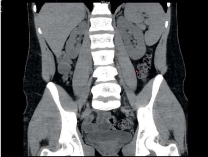
Рисунок 1. Компьютерная томография почек с контрастированием (апрель 2022 г.)
Figure 1. Computed tomography of the kidneys with contrast (April 2022)
Based on the anamnesis data, CT with contrast, and an increase in BP to 150/90 mm Hg, it was decided to continue antihypertensive therapy including enalapril 20 mg × 2 times/day and nifedipine 15 mg × 2 times/day.
On June 30, 2022, the child was consulted by a nephrologist at FSBI of the Russian Children’s Clinical Hospital (Moscow), where the diagnosis of “Secondary arterial hypertension, unspecified” was established. As a result, antihypertensive therapy was replaced: enalapril was discontinued, amlodipine was prescribed (10 mg × 2 times a day), and if BP increased to more than 130/90 mm Hg, sublingual nifedipine (10 mg) was additionally recommended.
On July 20, 2022, an online consultation was conducted via the telemedicine system; the consultant was Doctor of Medical Sciences, Professor, Head of the Nephrology Department of the FSCC of the FMBAR (Moscow) Nurali Zoirovich Zokirov. Hospitalization in the nephrology department of the FMBAR (Moscow) was recommended with a diagnosis: “Secondary arterial hypertension (renovascular?)”. The concomitant diagnosis was “Hypoplasia of the right kidney. Bilateral focal nephrosclerosis”. However, the boy was not hospitalized due to family circumstances.
In December 2022, he was examined and treated at the SBI of the Ryazan Region “City Clinical Hospital No. 11” (Ryazan). Upon admission, BP was 150/100–180/120 mm Hg.
The CBT and biochemical blood test were without pathology.
Hormonal profile was as follows: thyroid-stimulating hormone 1.2 μIU/ml (norm 0.3–4.2 μIU/ml), free thyroxine (T4) 19.2 pmol/l (norm 12.6–21.0 pmol/l), aldosterone (after walking) 548 pmol/l (norm 190–830 pmol/l).
The urine test according to Zimnitsky indicated the following indices: 2100 ml drunk, 975 ml daytime diuresis, 520 ml nighttime diuresis, and specific gravity of 1002–1018.
Upon daily urine volume of 2200 ml, daily proteinuria was 0 g/day, and glucosuria was 0.
ECG (on December 20, 2022) showed bradycardia and impaired repolarization processes. During echocardiography, the heart chambers were not enlarged in size, global contractility of the left ventricle was not impaired (ejection fraction was 67%). Mitral valve prolapse was of the 1st degree, mitral regurgitation was insignificant.
Ultrasound of the kidneys and bladder before and after urination on December 26, 2022 showed the following: kidney contours were smooth. RD was 95 × 45 mm, echogenicity of the parenchymatous layer was unchanged, before urination calyces were 3 mm, the renal pelvis was 6 mm; after urination the renal pelvis was 5 mm, no stones or space-occupying formations were detected. RS was 112 × 50 mm, echogenicity of the parenchymatous layer was unchanged, before urination calyces were 3 mm, the renal pelvis was 6 mm, after urination the renal pelvis was 5 mm, no stones or space-occupying formations were detected.
The DUSG of the renal vessels on December 23, 2022 revealed tortuosity of the renal arteries, which was more pronounced on the right. The DUSG of the vessels of the head and neck found moderate tortuosity of the vertebral artery in the bone canal on the right, and smooth tortuosity of the vertebral artery in the bone canal on the left.
Based on laboratory and instrumental studies, the following antihypertensive therapy was prescribed: enalapril 10 mg × 2 times a day, amlodipine 5 mg × 2 times a day, and bisoprolol 2.5 mg × 1 time per day. During the treatment, BP decreased to 115/70 mm Hg, and once dropped to 90/70 mm Hg. Due to the identified facts, it was decided to discontinue bisoprolol, afterwards BP was within the range of 130–120/80–70 mm Hg.
. On February 22, 2023, the child was admitted to the State Budgetary Healthcare Institution “Z.A. Bashlyaeva Children City Clinical Hospital” (Moscow) for examination and treatment.
During daily BP monitoring (on February 23, 2023), stable systolic AH was detected during the waking period.
An ultrasound scan (on February 23, 2023) of the renal arteries, kidneys, and adrenal glands revealed no pathology.
ECG (on February 23, 2023) showed an impairment of intraventricular conduction.
According to echocardiography (on February 24, 2023), the heart cavities and myocardial thickness were within normal limits, the valve apparatus was normal, the left ventricular ejection fraction was 72%, and bradycardia was recorded.
Based on the results of laboratory and instrumental studies, the diagnosis was “Stable arterial hypertension of the 1st degree”. Antihypertensive therapy was adjusted: cardosal 10 mg × 1 time in the morning and amlodipine 5 mg × 2 times a day were prescribed. As a result of treatment, BP was 120–130/70 mm Hg.
After discharge, the child continued to receive the prescribed treatment. However, during dynamic observation, BP periodically decreased to 100/70 mm Hg, so the dose of amlodipine was reduced to 5 mg × 1 time per day.
Further, the child was recommended to be monitored by a local pediatrician at his place of residence.
On December 19, 2023, the child was admitted to the pediatric department of the SBI of the Ryazan Region “City Clinical Hospital No. 11” (Ryazan) for a follow-up examination.
On admission, the patient was in a moderate condition, felt well, and had a BP of 120/70 mm Hg on the right arm.
In the CBT and GUT, no pathologies were detected.
The urine test according to Zimnitsky was as follows: 2150 ml drunk, 300 ml DD, 410 ml ND, and specific gravity of 1003–1027.
EchoCG on December 20, 2023 showed grade 1 mitral valve prolapse and grade 1 mitral regurgitation.
Ultrasound of the kidneys and bladder before and after urination on December 20, 2023 revealed that kidney contours were smooth. RD was of 100 × 44 mm, echogenicity of the parenchymal layer was unchanged, before urination the pelvis was 10 mm, after urination it was without dynamics, no stones or space-occupying formations were detected. RS was 106 × 46 mm, echogenicity of the parenchymal layer was unchanged, before urination the pelvis was 8 mm, after urination the pelvis was without dynamics, no stones or space-occupying formations were detected. The bladder was without pathology.
During the USDG of the renal vessels on December 20, 2023, a scattered type of structure of the renal arteries was revealed; tortuosity of the renal arteries was recorded, which was more pronounced on the right.
Antihypertensive therapy was selected, including cardosal 10 mg in the morning and amlodipine 5 mg in the morning. During dynamic observation in the department, the patient’s BP increased to 140/85–90 mm Hg.
Further, the child was recommended to continue antihypertensive therapy: cardosal 10 mg in the morning, long-term; amlodipine 5 mg in the morning but in case of an increase in BP > 130/80 mm Hg, an evening dose of 2.5–5 mg should be added; mexidol 1 tablet 2 times a day for 1 month. In addition, a consultation with a vascular surgeon was recommended, since pathology of the renal arteries of the right kidney was detected.
Currently, the child’s condition is stable, and dynamic observation is underway.
This clinical case demonstrates that secondary AH may be accompanied by absence of complaints and clinical symptoms for a long time. Regular monitoring of BP in children and adolescents, not only at specialist appointments, but also during preventive examinations by a pediatrician, is a requirement for early diagnosis of the disease and selection of examination and treatment tactics.
1. Hypertension Endangers Global Human Health. World Health Organization (WHO). 2022.
1. Aleksandrov A.A., Kisliak O.A., Leontyeva I.V. Clinical guidelines on arterial hypertension diagnosis, treatment and prevention in children and adolescents. Systemic Hypertension. 2020;17(2):7-35. (In Russ.) https://doi.org/10.26442/2075082x.2020.2.200126
2. Razumovsky A.Y., Alkhasov A.B., Bataev S.M., Abdurazakov M.A. Surgical treatment of vasorenal hypertension in children. Russian Journal of Pediatric Surgery, Anesthesia and Intensive Care. 2018;8(1):36-43. (In Russ.) https://doi.org/10.30946/2219406120188136-43
3. Latypov T.H., Chekmareva I.A., Kazantseva G.P., Deev R.V. Structure and ultrastricture of kidney tissue with the Good-pasture’s syndrome: a clinical case. Science of the young (Eruditio Juvenium). 2018;6(3):405-413. (In Russ.) https://doi.org/10.23888/HMj201863405-413
4. Bushueva E.V., Gerasimova L.I., Dianova T.I., Ivanova O.N., Smirnova E.I. Dynamics of blood pressure indicators in children and adolescents over two decades (1999-2022). Modern problems of science and education. 2022;6-1. (In Russ.). https://doi.org/10.17513/spno.32252
5. Bekezin V.V. Arterial hypertension in children and adolescents. Smolensk medical Almanac. 2016;(3):192-209. (In Russ.). eLIBRARY ID: 27421509 EDN: xCBRqF
6. Luma GB, Spiotta RT. Hypertension in children and adolescents. Am Fam Physician. 2006;73(9):1558-1568. PMID: 16719248
7. Chazova I.E., Chikhladze N.M., Blinova N.V., Belaya Zh.E., Danilov N.M., et al. Eurasian clinical guidelines for the diagnosis and treatment of secondary (symptomatic) forms of arterial hypertension (2022). Eurasian heart journal. 2023;(1):6-65. (In Russ.) https://doi.org/10.38109/2225-1685-2023-1-6-65
8. Raina R, Mahajan Z, Sharma A, Chakraborty R, Mahajan S, et al. Hypertensive Crisis in Pediatric Patients: An Overview. Front Pediatr. 2020;8:588911. https://doi.org/10.3389/fped.2020.588911
9. Nikolaev N.A. Evidence-based hypertensiology: quantitative assessment of the results of antihypertensive therapy. Moscow: Academy of Natural Sciences. 2008. (In Russ.).
10. Spagnolo A, Giussani M, Ambruzzi AM, Bianchetti M, Maringhini S, et al. Focus on prevention, diagnosis and treatment of hypertension in children and adolescents. Ital J Pediatr. 2013;39:20. https://doi.org/10.1186/1824-7288-39-20
11. Anyaegbu EI, Dharnidharka VR. Hypertension in the teenager. Pediatr Clin North Am. 2014;61(1):131-151. https://doi.org/10.1016/j.pcl.2013.09.011
12. Seleznev S.V., Yаkushin S.S., Mylnikov P.Y., Tranova Y.S., Shchul'kin A.V., et al. Therapeutic Drug Monitoring in Uncontrolled Arterial Hypertension: Result of the Pilot Part of Study. I.P. Pavlov Russian Medical Biological Herald. 2023;31(2):195-202. https://doi.org/10.17816/PAVLOVj119880
Natalya A. Belykh. Dr.Sci. (Med.), Docent, Head of the Department of Polyclinic Pediatrics with the Course of Pediatrics of the Faculty of Additional Professional Education
Ryazan
Authors declares no conflict of interest.
Yulia S. Kucheryavenkova, student
Ryazan
Authors declares no conflict of interest.
Inna V. Piznyur, assistant of the Department of Polyclinic Pediatrics with the Course of Pediatrics of the Faculty of Additional Professional Education
Ryazan
Authors declares no conflict of interest.
Alla Yu. Lashko, is a pediatrician
Ryazan
Authors declares no conflict of interest.
Nataliya A. Anikeeva, MD, PhD, Assistant Professor of the Department of Polyclinic Pediatrics with the Course of Pediatrics of the Faculty of Additional Professional Education
Ryazan
Authors declares no conflict of interest.
Belykh N.A., Kucheryavenkova Y.S., Piznyur I.V., Lashkov A.Yu., Anikeeva N.A. Clinical case of secondary arterial hypertension in the practice of a pediatric. Medical Herald of the South of Russia. 2024;15(4):66-71. (In Russ.) https://doi.org/10.21886/2219-8075-2024-15-4-66-71
29, Nakhichevansky Lane, Rostov-on-Don, 344002
Rostov State Medical University
Тel.: +79185710558
e-mail: rostgmu-journal@rambler.ru


