


Contents
Scroll to:
https://doi.org/10.21886/2219-8075-2022-13-2-80-85
Scroll to:
A case of early prenatal diagnosis of diastrophic dysplasia at 13 weeks 2 days of gestation is described. During ultrasound screening of the first trimester, fetal changes characteristic of this form of systemic skeletal dysplasia were revealed – micrognathia, micromelia, flexion contracture in the elbow and knee joints, pathognomonic abduction of the thumb of the hands and feet for diastrophic dysplasia (hitchhiker thumb), varus deviation and postaxial polydactyly of the feet, in combination with by a significant increase in the nuchal translucency. Pregnancy was interrupted for medical reasons. The possibilities of ultrasound diagnostics of diastrophic dysplasia by two- and three-dimensional ultrasound using are shown.
Tyo S.A. A case of prenatal diagnosis of diastrophic dysplasia in the 1st trimester of pregnancy. Medical Herald of the South of Russia. 2022;13(2):80-85. (In Russ.) https://doi.org/10.21886/2219-8075-2022-13-2-80-85
Diastrophic dysplasia (DD), OMIM 222600 (diastrophic dwarfism syndrome) is a rare systemic skeletal dysplasia (SSD) with an autosomal recessive type of inheritance, the main manifestations of which are shortening of the limbs by the type of micromelia or rhizomelia, clubfoot, deformity of the hands and feet, multiple flexor contractures and scoliosis, deformation and thickening of the auricles, and cleft palate in 25% of cases. It does not apply to lethal skeletal dysplasia, as well as it is not combined with cognitive disorders. In addition to family forms, sporadic cases also occur [1][2].
For the first time, the term “diastrophic” was borrowed by Lamy and Maroteaux in 1960 from geology: diastrophism is tectonic movements and deformation of the Earth's crust [3]. For the first time, the authors identified DD as a separate nosological unit, which was previously regarded as a variant of other SSDs, in particular achondroplasia with clubfoot, multiple congenital arthrogryposis, or multiple congenital malformations and counterattacks [4][5].
The frequency of DD occurrence is 1 newborn case in 100,000, and in Finland, its maximum spread is noted (0.3 cases per 10,000 newborns), which is due to the high concentration of the IVS1+2T>C mutation in the SLC26A2 gene [1][6]. This is probably why the gene was first mapped in the Finnish population in 1990, when children with DD from 13 families were specifically examined [7].
Further studies have confirmed that the occurrence of DD is associated with a homozygous or complex heterozygous mutation in the SLC26A2 gene (DTDST) on the long arm of chromosome 5 (5q32-q33.1), which encodes a transmembrane protein that transports sulfates to chondrocytes and fibroblasts to maintain adequate sulfation of proteoglycans. Violation of this process prevents endochondral ossification and, consequently, leads to abnormal growth and remodeling of bone and cartilage tissues. When one gene was affected, a different degree of mutation influence on the process of sulfate transport was revealed; probably, this process in combination with other factors leads to the development of four types of chondrodysplasia with different clinical phenotypes and prognoses. These include type IB achondrogenesis and type II atelosteogenesis, which are lethal forms, and non-lethal forms such as DD and recessive multiple epiphyseal dysplasia [8, 9].
The first publication on the possibilities of echography in the diagnosis of DD dates back to 1983, in which Finnish authors cite a case of prenatal diagnosis of this chondrodysplasia at 16 weeks of pregnancy in a patient at risk (a previous child with DD), confirmed at 19 weeks by fetoscopy and X-ray examination [10].
Despite the fact that the clinical core of DD is extremely pronounced and most pregnant women undergo screening of trimesters I and II in specialized institutions and trained specialists, there are not so many publications on this topic, including in domestic journals. At the same time, most of them are devoted to the possibilities of prenatal diagnosis of this skeletal dysplasia in the first trimester, which increases their value, but the last domestic publication dates back to 2015 [11-15][6][16].
The authors of this particular study present their own observation of early prenatal diagnosis of DD at 13 weeks of pregnancy.
A pregnant patient, 34 years old, the 4th pregnancy, there were 3 abortions (because of non-developing pregnancies up to 12 weeks). The spouses were healthy, the family history was not burdened, the marriage was unrelated, they had no professional harms.
During the ultrasound first trimester screening (at the place of pregnancy observation), micrognathia and shortening of the limbs were revealed. The patient was sent for examination in order to clarify the diagnosis with the medical conclusion “signs of skeletal dysplasia”.
The ultrasound examination was performed by means of using the Accuvix-A30 device (Samsung Madison) with a volumetric convexic sensor V 4-8 and a volumetric recto-vaginal sensor V 5-9.
This ultrasound examination revealed one living fetus in the uterine cavity (Figure 1). The coccygeal-parietal size was 65 mm (12 weeks 6 days), biparietal size — 24 mm, head circumference — 85 mm, abdominal circumference — 74 mm, hip length — 5 mm (less than 5 percentile), heartbeat 151 beats/min. The thickness of nuchal translucency (NT scan) was 6.8 mm, nasal bones were visualized, the venous duct was 1.08, reverse blood flow was not determined, there was no tricuspid regurgitation (Figure 2).

Рисунок 1. Продольное сканирование плода. 1 — микрогнатия; 2 — увеличенная толщина воротникового пространства; 3 — деформированная стопа с постаксиальной полидактилией и отведением большого пальца.
Figure. 1. Longitudinal scanning of the fetus. 1 – micrognathia; 2 – increased thickness of nuchal translucency; 3 – deformed foot with postaxial polydactyly and thumb retraction.

Рисунок 2. Профиль плода (срединно-сагиттальный срез).
Figure. 2. Profile of the fetus with micrognathia (mid-sagittal section).
The shape of the head, the bones of the skull, and the structure of the main structures of the brain were without features (Figure 3). No obvious hypoplasia of the chest was detected. A four-chamber section and a section through 3 vessels (V-sign) were without features as well (Figure 4). The anterior abdominal wall, abdominal organs, kidney area, and bladder were without features. The spine was not changed.

Рисунок 3. Поперечное сечение головы (А) и живота (Б).
Figure. 3. Axial section of the head (A) and abdomen (B).

Рисунок 4. Четырёхкамерный срез сердца (А) и срез через 3 сосуда — V-sygn (Б) в режиме ЭДК.
Figure. 4. Four-chamber section of the heart (A) and a three-vessels view – V-sign (B) in PD mode.
The structure of the chorion was not changed, the umbilical cord had 3 vessels, attachment to the chorion was without features.
While assessing facial structures, pronounced micrognathia was noted without displacement of the auricles downwards and signs of cleft of the face, in particular the hard palate.
Upper and lower limbs were with signs of flexion contracture in the elbow and knee joints, as well as the significant shortening of long tubular bones by the type of micromelia (Figures 5, 6).

Рисунок 5. Верхняя конечность — микромелия, сгибательная контрактура локтевого сустава, большой палец «путешественника автостопом».
Figure. 5. Upper limb – micromelia, flexion contracture of the elbow joint, thumb of the “hitchhiker".


Рисунок 6. Нижние конечности. А. Сгибательная контрактура коленных суставов, микромелия. 1 — бедра; 2 — голени. Б. Девиация стоп. 1 — левая стопа; 2 — правая стопа.
Figure. 6. Lower extremities. A. Flexion contracture of knee joints, micromelia. 1 – hips; 2 – lower legs. B. Deviation of the feet. 1 – left foot; 2 – right foot.
The hands had characteristic “hitchhiker” thumbs (Figure 7).
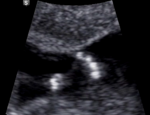
Рисунок 7. Кисть с характерным радиальным отведением большого пальца — признак “hitchhiker thumb”.
Figure. 7. A brush with a characteristic radial thumb retraction is a sign of a "hitchhiker thumb".
The angle of the foot placement was changed according to the type of varus deviation, and postaxial polydactyly of both feet, an increase in the size of the thumbs, and their characteristic deviation to the side were also determined (Figure 8).
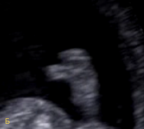
Рисунок 8. Стопа. А. Варусная девиация, постаксиальная полидактилия, отведение большого пальца. Б. Отведение большого пальца.
Figure. 8. Foot. A. Varus deviation, postaxial polydactyly, thumb abduction. B. Thumb abduction.
During the three-dimensional (3D) reconstruction, the pathological changes in the fetus, previously detected in 2D mode, were fully confirmed (Figure 9).
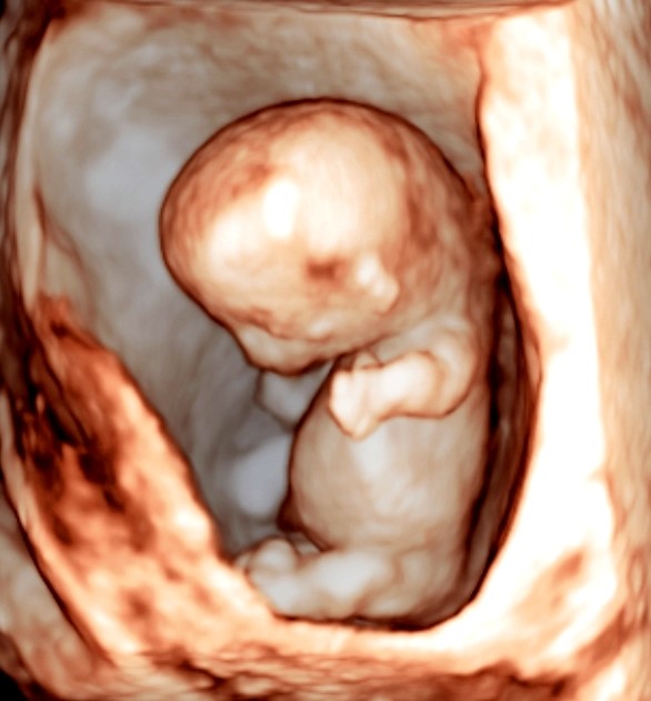
Рисунок 9. Трёхмерное изображение плода в режиме поверхностной реконструкции.
Figure. 9. Three-dimensional image of the fetus in the mode of surface reconstruction
The revealed complex of ultrasound signs fully corresponded to the clinical core of DD as the main diagnostic hypothesis for such an echographic picture.
The pregnancy was terminated for medical reasons. Targeted sequencing of abortus and a married couple with subsequent medical and genetic counseling was recommended.
The complexity of prenatal diagnosis of SSDs is due to the fact that their spectrum is quite wide, as well as the variability of phenotypic manifestations and the timing of prenatal manifestation. Nevertheless, most of them have characteristic pathognomonic signs or a combination of changes that facilitates the diagnostic search and identification of the nosological form [17].
With careful adherence to the methodology of ultrasound first trimester screening, a wide range of SSDs is also available for early prenatal diagnosis [18][19].
Currently, echography still continues to be the main tool for the primary prenatal diagnosis of SSD. The basic two-dimensional visualization mode has been successfully supplemented with various options for reconstructing the three-dimensional image of the fetus. In particular, surface reconstruction and skeletal mode allow improving the detail and visual perception of the revealed changes [12][16]. In the presented case, the three-dimensional image of the fetus in the mode of surface reconstruction fully reflected the essence of the pathological changes characteristic of DD, including its pathognomonic sign – radial abduction of the thumbs of the hands (Figure 10). A very important point that increases the effectiveness of early detection and the possibility of differential diagnosis of SSD is a detailed assessment of the hands and feet [19].
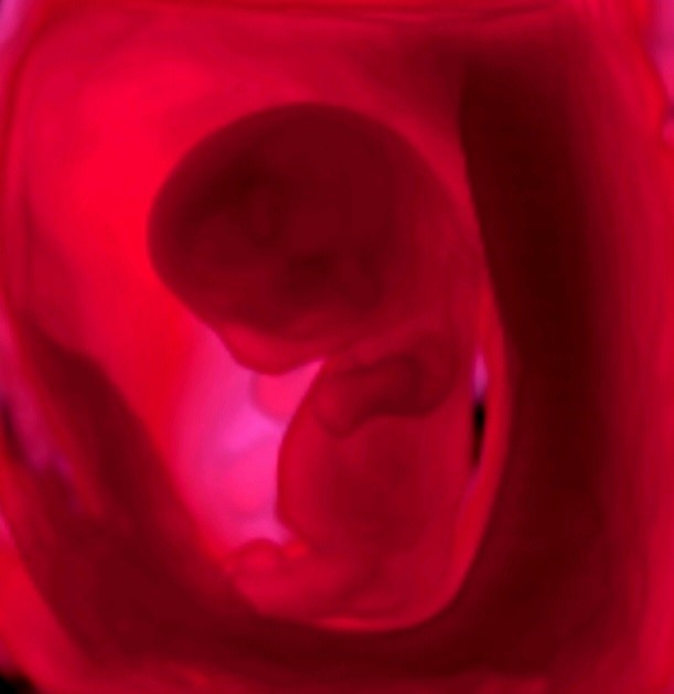
Рисунок 10. Трёхмерное изображение плода в режиме Fetal Realistic Vue.
Figure. 10. Three-dimensional image of the fetus in Fetal Realistic Vue mode.
It should be noted that the increase in the thickness of the collar space revealed in this case refers to non-specific early ultrasound markers of various forms of SSD [20, 21]. This sign signals the need for a more thorough assessment of the fetal skeleton, with the mandatory use of transvaginal access.
Of course, in order to fully verify cases of prenatally detected SSDs, a comprehensive study of abortion is necessary, which should include radiological diagnostics — X-ray (recommended minimum), CT or MRI (if necessary), pathoanatomic, histological and genetic studies, followed by medical and genetic counseling of a married couple before planning the next pregnancy. However, as practice and the presented case show, after the termination of pregnancy, the full range of verifying diagnostic measures is not carried out and only ultrasound data remain for medical and genetic counseling. Therefore, while detecting SSDs, a prenatal diagnostic specialist needs to record each detected ultrasound sign as accurately as possible on echograms and video clips, which can facilitate subsequent consultation by a geneticist.
Despite the fact that after the termination of pregnancy, it was impossible to obtain information about conducting any additional studies of both abortus and a married couple, the identified set of changes, including the pathognomonic sign of radial abduction of the thumb, allows stating with sufficient confidence that the authors of this study faced the ultrasound DD semiotics.
Thus, the presented observation confirms the possibilities of early prenatal diagnosis of DD within the framework of ultrasound screening of the first trimester of pregnancy, and 3D echography improves the visual perception of the detected changes in the fetus, making them as realistic as possible and increasing the accuracy of determining the nosological form of SSD.
1. Medvedev M.V. Prenatal'naja jehografija. Differencial'nyj diagnoz i prognoz. 4-e izd., dop., perer. Moscow: Real Tajm; 2016. (In Russ.)
2. Kozlova S.I., Demikova N.S. Nasledstvennye sindromy i medikogeneticheskoe konsul'tirovanie. 3-e izdanie, pererabotannoe i dopolnennoe. Moscow: T-vo nauchnyh izdanij KMK; Avtorskaja akademija; 2007. (In Russ.)
3. Lamy M, Maroteaux P. Diastrophic nanism. Presse Med. 1960;68:1977-80. (in French). PMID: 13758600.
4. Mau H. Wesen und Bedeutung der enchondralen Dysostosen. Stuttgart: Georg Thieme Verlag (pub.); 1958.
5. Kite j.H. The Clubfoot. New York: Grune and Stratton (pub.); 1964.
6. Honório jC, Bruns RF, Gründtner LF, Raskin S, Ferrari LP, et al. Diastrophic dysplasia: prenatal diagnosis and review of the literature. Sao Paulo Med J. 2013;131(2):127-32. DOI: 10.1590/s1516-31802013000100024.
7. Hästbacka j, Kaitila I, Sistonen P, de la Chapelle A. Diastrophic dysplasia gene maps to the distal long arm of chromosome 5. Proc Natl Acad Sci U S A. 1990;87(20):8056-9. DOI: 10.1073/pnas.87.20.8056.
8. Rossi A, Superti-Furga A. Mutations in the diastrophic dysplasia sulfate transporter (DTDST) gene (SLC26A2): 22 novel mutations, mutation review, associated skeletal phenotypes, and diagnostic relevance. Hum Mutat. 2001;17(3):159-71. DOI: 10.1002/humu.1.
9. Karniski LP. Mutations in the diastrophic dysplasia sulfate transporter (DTDST) gene: correlation between sulfate transport activity and chondrodysplasia phenotype. Hum Mol Genet. 2001;10(14):1485-90. DOI: 10.1093/hmg/10.14.1485.
10. Kaitila I, Ammälä P, Karjalainen O, Liukkonen S, Rapola j. Early prenatal detection of diastrophic dysplasia. Prenat Diagn. 1983;3(3):237-44. DOI: 10.1002/pd.1970030309.
11. Pribushenja, O.V., Novikov I.V., Kovalev S.I., Krapiva G.A., Plevako T.A., et al. Vozmozhnosti prenatal'noj diagnostiki distroficheskoj displazii v I trimestre beremennosti. Prenatal Diagnosis. 2005;4(1):56-61. (In Russ.).
12. Badigova E.A. Sluchaj rannej prenatal'noj diagnostiki diastoficheskoj displazii. Prenatal Diagnosis. 2010;9(1):84-86. (In Russ.). eLIBRARY ID: 22829559
13. Rusanova O.K., Grjashhenko V.N., Nikolaeva O.P., Lakomskaja E.V. Prenatal'naja diagnostika skeletnyh displazij: ahondrogenez (tip IV) i diastroficheskaja displazija. Prenatal Diagnosis. 2011;10(1):56-61. (In Russ). eLIBRARY ID: 22678416
14. Novikova I.V., Lazarevich A.A., Solovyeva I.V., Venchikova N.A., Marachovskaya E.I. Prenatal ultrasound diagnosis of diastrophic dysplasia at 12 weeks of gestation. Prenatal Diagnosis. 2015;14(2):130-136. (In Russ.). eLIBRARY ID: 23731034
15. Grammatikova O.A., Ljutaja E.D., Starikova T.ju., Osadshaja V.N. Prenatal'naja diagnostika diastroficheskoj displazii real'no mozhet byt' osushhestvlena pri skriningovom ul'trazvukovom issledovanii v 11-14 nedel' beremennosti. Prenatal Diagnosis. 2015;14(3):269- 271. (In Russ.). eLIBRARY ID: 24114359
16. de Souza Lima T, Ferreira BG, Loureiro Souza Cw, Batista IBC, Araujo júnior E, et al. Prenatal diagnosis of diastrophic dysplasia in the second trimester of pregnancy: Two- and three- dimensional ultrasonographic findings. Turk J Obstet Gynecol. 2021;18(3):258- 263. DOI: 10.4274/tjod.galenos.2021.35033.
17. Medvedev M.V. Prenatal'naja jehografija. Differencial'nyj diagnoz i prognoz. 2-e izd., perer. M.: Real Tajm; 2009. (In Russ.).
18. Shevchenko E.A. Prenatal'naja ul'trazvukovaja diagnostika skeletnyh displazij v 11 – 14 nedel' beremennosti. Prenatal Diagnosis. 2007;6(2):114 – 122. (In Russ.).
19. Lazarevich A.A. Ul'trazvukovye differencial'nye priznaki sistemnyh skeletnyh displazij u plodov 1 trimestra beremennosti i jetapy prenatal'noj diagnostiki. Medicine: theory and practice. 2019;4(S):299-300. (In Russ.). eLIBRARY ID: 39199171
20. Ngo C, Viot G, Aubry MC, Tsatsaris V, Grange G, et al. Firsttrimester ultrasound diagnosis of skeletal dysplasia associated with increased nuchal translucency thickness. Ultrasound Obstet Gynecol. 2007;30(2):221-6. DOI: 10.1002/uog.4028.
21. Nikolaides K. Ul'trazvukovoe issledovanie v 11 – 13+6 nedel' beremennosti. Per. s angl. SPb.: ID «Petropolis»; 2007. (In Russ.)
Sergey A. Tyo, Cand. Sci. (Med.), ultrasound diagnostics in obstetrics and gynecology
Moscow
Tyo S.A. A case of prenatal diagnosis of diastrophic dysplasia in the 1st trimester of pregnancy. Medical Herald of the South of Russia. 2022;13(2):80-85. (In Russ.) https://doi.org/10.21886/2219-8075-2022-13-2-80-85
29, Nakhichevansky Lane, Rostov-on-Don, 344002
Rostov State Medical University
Тel.: +79185710558
e-mail: rostgmu-journal@rambler.ru


