


Contents
Scroll to:
https://doi.org/10.21886/2219-8075-2021-12-3-78-85
Scroll to:
Objective: To perform a comparative analysis of the linear dimensions of the facial skull that are the most signifi cant in determining asymmetry in persons of diff erent sex on spiral computed tomograms (SCT).
Materials and Methods: The CT scan of 104 people of both sexes (women – 52%, n = 54, men – 48%, n = 50) were studied. Measurements were made using a standard digital ruler of a computer tomograph workstation, and the asymmetry of the skulls was assessed using the “fan” method. The results were processed using the Excel program.
Results: In the male and female series of SCT, according to the size of the intervals of sigma of linear dimensions of the facial skull, the occurrence rate of asymmetry of varying severity was determined.
Conclusion: In the male series of SCT, right-sided asymmetry of the linear dimensions of the facial skull prevails in all “fans”, in the female – right-sided asymmetry in the upper and lateral “fans”, but left -sided asymmetry in the lower “fan”. Statistically signifi cant linear dimensions of the facial skull were revealed to determine the severity of asymmetry in both sexes. In the upper “fan”, these are the distances from the nasion to the zygomaxilar (N-Zm), from the nasion to the frontonazale (N-Fn), prevailing on the right in both sexes. In the lower “fan”– the distance from the supraspinal to the zygomaxilar (Ss-Zm), and in the lateral “fan” – the distance from the zygomaxilar to the frontonasal (Zm-Fn), prevailing on the right in both sexes. An insignifi cant or physiological and moderate degree of asymmetry in the size of the facial skull depending on gender was revealed, which did not require correction.
Shepetyuk M.G., Kaplunova O.A., Shepetyuk M.G., Suhanova O.P., Blinov I.M. Sexual features of facial skull asymmetry according to spiral computed tomography. Medical Herald of the South of Russia. 2021;12(3):78-85. (In Russ.) https://doi.org/10.21886/2219-8075-2021-12-3-78-85
The fact of facial asymmetry in people is generally acknowledged. One of the reasons for this asymmetry is an uneven assembly of skull bones elements [1]. For many years, traditionally, the main method of analysis of the morphology of the facial skull and diagnostics of facial bones deformation was X-ray imaging. However, it is difficult to evaluate the asymmetry of the facial skull because of numerous overlapping anatomic structures [2]. Computer-assisted craniometry is a vast field of study because tomographic studies of high resolution have become acknowledged standards of diagnostic [3]. The results of craniometry of macerated skulls, X-ray images, and spiral computed tomography show that spiral computed tomography (SCT) can be useful and alternative to a regular X-ray [4]. The results of the measurements obtained from SCT are precise and comparable with the results of anthropometric analysis of a macerated skull [5][6][7], which expands the clinical application of SCT [8][9].
It is known that the individual shape of the face determines the physiological asymmetry of the facial skull [10]. Along with this, such asymmetry of bone structures of the skull in modern people is understudied, in particular, in a sex-related aspect [4][11]. Thus, a comparative analysis of craniometric parameters in persons of both sexes residing in the Rostov Region is acute and relevant.
The study aimed to evaluate the degree of expression of asymmetry of linear sizes of the facial skull that are the most significant in determining asymmetry in persons of both sexes by the results of SCT.
SCT scans of 104 people of both sexes (52% of women (n = 54) and 48% of men (n = 50)) were studied. SCT was performed at the facilities of the department of magnetic resonance imaging and computed tomography at the Rostov State Medical University in patients with suspected vascular pathology of the brain.
Criteria of inclusion:
Criteria of exclusion:
For the analysis of the obtained results, the authors used axial, MPR (multiplanar reconstruction), and SSD (surface shadow density) reconstructions of the skull in different projections. The measurements were made using a standard digital scale of the operating station of a computed tomograph.
The shapes of the brain and facial skull were determined by the dimensions of a cephalic index [12]. A “fan” method was used in SCT scanning [13][14] in the upper, lower, and lateral fans for the measurement of the distance from standard points nasion, supraspinal, and zygomaxilar to non-standard points from each side. This method included the study of 27 linear dimensions of the facial skull from each side.
Processing of the statistical data was performed using the Excel software package recommended for the statistical analysis of medical-biological data. For each studied parameter, the authors calculated the sample mean value (M) and the standard error of the mean (m). The significance of the differences in the mean values was evaluated using Student’s parametric criterion for normal distribution of the initial data. The differences between the groups of parameters were considered significant at p < 0.05.
According to the recommendations of Efimova et al. [15], during the calculation of the coefficient of variation (kv=δ/M – the ratio of the standard deviation of linear dimensions to the arithmetic mean of these dimensions), kv < 10 % was considered to be a weak expression, 10% < kv < 25% was considered to be a moderate expression, and kv > 25% – a high degree of expression. The method of Gaivoronskiy et al. [10] was used to evaluate mean linear dimensions of the facial skull on the right and left, to calculate the differences between them in the sigmas, and the degree of expression of asymmetry of the facial skull within the sigmas 1, 2, and 3.
During the distribution of material depending on the sex, SCT in men and women revealed significant differences in 27 linear dimensions of the facial skull in the upper, lower, and lateral “fans” on both sides (Table 1).
SCT revealed right-sided asymmetry of linear dimensions of the facial skull in all “fans” in men. In women, SCT revealed right-sided asymmetry in the upper and lateral “fans” and left-sided asymmetry in the lower “fan”.
In the majority of cases, the coefficient of variation of the studied linear dimensions of the facial skull did not exceed 10–20%, which indicated the weak and moderate variation of the parameters of each dimension and their consistency.
In the groups of males and females, SCT images were used to determine the occurrence rate of asymmetries by the intervals of sigmas of these dimensions. In the series of SCT of men (Fig. 1), in the upper, lower, and lateral “fans”, the differences in the parameters on both sides did not exceed 1 ϭ in 67.6%, 74.9%, and 64.3% of cases, respectively; they were within 2 ϭ (in 25.7%, 15.6%, and 30.3% of cases, respectively); and they were within 3 ϭ (in 6.8%, in 8.6%, and 5.4% of cases, respectively). In the series of SCT of women (Fig. 2), in the upper, lower, and lateral “fans”, the differences on both sides did not exceed 1 ϭ in 68.8%, 63.8%, and 65.0% of cases; the differences were within 2 ϭ (in 26.3%, 32.5%, and 32.8% of cases, respectively); and they were within 3 ϭ (in 4.9%, 3.7%, and 2.2% of cases, respectively).
Table 1
The linear dimensions of the facial skull with different degrees of asymmetry in persons of both sexes, p < 0.05, M ± m (mm), kv (%), σ (%)
|
Linear dimension |
Men |
Women |
|
||||||
|
Indicator values to the right (D) or left (S) in mm |
The frequency of occurrence of size asymmetry within 1σ-2 σ-3σ (%) |
Kv size (%)
|
Indicator values to the right (D) or left (S) in mm |
The frequency of occurrence of size asymmetry within 1σ-2 σ-3σ (%) |
Kv size (%)
|
|
|||
|
Upper fan |
|
||||||||
|
N-Ft |
S |
46.8±4.6 |
64-29-7 |
9.9 |
47.9±6.4 |
75-21-4 |
13.4 |
||
|
D |
47.4±4.9 |
71-22-7 |
10.3 |
48.3±6.6 |
75-19-6 |
13.7 |
|||
|
N-Fmt |
S |
48.3±5.5 |
67-27-7 |
11.3 |
48.7±6.6 |
69-25-6 |
13.6 |
||
|
D |
48.9±5.7 |
71-24-4 |
11.6 |
49.2±6.8 |
75-19-6 |
13.9 |
|||
|
N-Da |
S |
15.0±3.1 |
62-33-4 |
20.3 |
15.8±4.1 |
63-35-2 |
26.2 |
||
|
D |
14.9±2.8 |
67-27-7 |
18.9 |
15.8±3.9 |
62-37-2 |
24.7 |
|||
|
N-Infr |
S |
30.5±4.3 |
58-38-4 |
14.2 |
30.9±4.6 |
69-27-4 |
14.9 |
||
|
D |
31.5±4.0 |
71-20-9 |
12.6 |
32.3±5.2 |
69-25-6 |
16.1 |
|||
|
N-Zm* |
S |
58.8±5.6 |
67-16-18 |
9.6 |
56.0±7.4 |
69-27-4 |
13.2 |
||
|
D |
59.0±5.6 |
64-18-18 |
9.5 |
57.2±7.5 |
69-27-4 |
13.1 |
|||
|
N-Fn* |
S |
5.1±1.0 |
69-18-13 |
20.3 |
6.1±1.8 |
65-29-6 |
30.0 |
||
|
D |
5.8±1.2 |
56-40-4 |
20.9 |
6.8±1.9 |
73-27-0 |
28.3 |
|||
|
N-max |
S |
26.5±4.1 |
67-22-11 |
15.5 |
25.0±3.9 |
56-37-8 |
15.7 |
||
|
D |
26.2±4.0 |
69-24-7 |
15.2 |
25.0±3.7 |
63-35-2 |
15.0 |
|||
|
N-ap.lat |
S |
36.3±4.5 |
76-16-9 |
12.4 |
37.5±5.8 |
69-25-6 |
15.5 |
||
|
D |
37.0±4.2 |
71-18-11 |
11.3 |
37.3±5.9 |
75-17-8 |
15.7 |
|||
|
N-ap inf |
S |
43.2±4.8 |
69-20-11 |
11.2 |
43.7±6.7 |
75-15-10 |
15.2 |
||
|
D |
43.5±4.7 |
69-18-13 |
10.8 |
43.8±6.7 |
75-19-6 |
15.4 |
|||
|
N-min |
S |
19.0±3.4 |
71-24-4 |
17.7 |
18.9±2.9 |
62-33-6 |
15.2 |
||
|
D |
18.8±3.1 |
73-22-4 |
16.7 |
19.2±2.9 |
65-29-6 |
15.0 |
|||
|
Lower fan |
|||||||||
|
Ss-Ft |
S |
72.5±11.3 |
84-9-7 |
15.5 |
73.6±8.6 |
67-27-6 |
11.6 |
||
|
D |
73.7±7.7 |
73-18-9 |
10.4 |
73.1±8.1 |
71-23-6 |
11.1 |
|||
|
Ss-Fmt |
S |
68.3±7.4 |
78-11-11 |
10.8 |
69.7±9.2 |
65-29-6 |
13.1 |
||
|
D |
68.2±7.1 |
79-9-13 |
10.4 |
69.2±9.2 |
69-25-6 |
13.3 |
|||
|
Ss-zm*
|
S |
46.0±5.4 |
67-16-18 |
11.8 |
42.8±4.5 |
71-25-4 |
10.6 |
||
|
D |
45.3±5.2 |
67-22-11 |
11.4 |
42.7±4.9 |
75-23-2 |
11.4 |
|||
|
Ss-Fn |
S |
44.1±4.9 |
78-4-18 |
11.0 |
43.8±6.9 |
63-35-2 |
15.7 |
||
|
D |
44.2±5.1 |
78-9-13 |
11.4 |
44.2±6.7 |
69-29-2 |
15.1 |
|||
|
Ss-Da
|
S |
42.4±4.6 |
71-16-13 |
10.9 |
42.2±6.2 |
69-27-4 |
14.8 |
||
|
D |
42.6±4.6 |
73-13-13 |
10.9 |
42.2±5.9 |
63-33-4 |
14.1 |
|||
|
Ss-min
|
S |
28.3±4.4 |
80-13-7 |
15.4 |
28.9±5.9 |
62-35-4 |
20.5 |
||
|
D |
28.1±4.3 |
80-13-7 |
15.3 |
28.6±6.2 |
71-25-4 |
21.6 |
|||
|
Ss-infr
|
S |
33.2±4.5 |
80-9-11 |
13.5 |
33.0±3.8 |
65-33-2 |
11.5 |
||
|
D |
33.3±4.0 |
80-9-11 |
11.9 |
32.9±4.1 |
71-25-4 |
12.3 |
|||
|
Ss-max
|
S |
22.8±4.3 |
18-13-9 |
18.7 |
23.3±4.6 |
65-31-4 |
19.5 |
||
|
D |
22.3±3.9 |
80-13-7 |
17.4 |
23.0±4.7 |
67-29-4 |
20.5 |
|||
|
Ss-ap. lat |
S |
14.8±2.1 |
67-20-13 |
14.1 |
14.8±2.9 |
67-31-2 |
19.6 |
||
|
D |
15.0±1.9 |
62-27-11 |
12.7 |
15.3±3.0 |
65-33-2 |
19.3 |
|||
|
Ss-ap. Inf. |
S |
6.5±1.7 |
84-9-7 |
25.8 |
6.7±1.6 |
56-38-6 |
24.7 |
||
|
D |
6.8±1.6 |
60-38-2 |
23.8 |
7.3±1.8 |
58-40-2 |
24.1 |
|||
|
Side fan |
|||||||||
|
Zm-infr |
S |
22.5±3.4 |
64-27-9 |
15.1 |
22.4±4.2 |
65-33-2 |
18.8 |
||
|
D |
22.2±3.5 |
64-24-11 |
15.6 |
23.4±5.1 |
67-31-2 |
21.7 |
|||
|
Zm-ap.lat |
S |
20.9±3.6 |
62-31-7 |
17.3 |
20.2±3.6 |
62-37-2 |
17.7 |
||
|
D |
21.2±4.1 |
64-27-9 |
19.3 |
20.8±4.1 |
52-44-4 |
19.6 |
|||
|
Zm-ap.inf |
S |
18.0±3.0 |
71-22-7 |
16.9 |
17.7±3.5 |
75-23-2 |
19.7 |
||
|
D |
18.1±2.9 |
64-29-7 |
16.1 |
18.2±3.3 |
73-21-6 |
18.4 |
|||
|
Zm-max |
S |
35.7±8.2 |
64-31-4 |
23.1 |
32.7±6.8 |
56-44-0 |
20.7 |
||
|
D |
35.8±8.0 |
58-36-7 |
22.4 |
33.5±7.1 |
63-37-0 |
21.3 |
|||
|
Zm-Da |
S |
29.3±4.3 |
69-24-7 |
14.6 |
29.6±4.8 |
67-29-4 |
16.3 |
||
|
D |
30.0±4.2 |
60-33-7 |
13.9 |
31.0±5.3 |
65-35-0 |
17.1 |
|||
|
Zm-min |
S |
40.4±6.2 |
69-22-9 |
15.4 |
38.1±5.7 |
69-27-4 |
14.8 |
||
|
D |
40.5±6.2 |
67-22-11 |
15.2 |
38.4±5.8 |
63-37-0 |
15.2 |
|||
|
Zm-Fn* |
S |
46.0±5.6 |
60-33-7 |
12.1 |
43.1±4.7 |
63-37-0 |
11.0 |
||
|
D |
46.3±5.5 |
62-24-13 |
11.9 |
44.3±5.3 |
67-27-6 |
11.9 |
|||
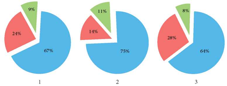
Fig. 1. Asymmetry of varying severity of the facial skull linear dimensions in men (%) within sigma 1 (blue), sigma 2 (red), and sigma 3 (green). 1 – upper “fan”, 2 – lower “fan”, 3 – lateral “fan”.
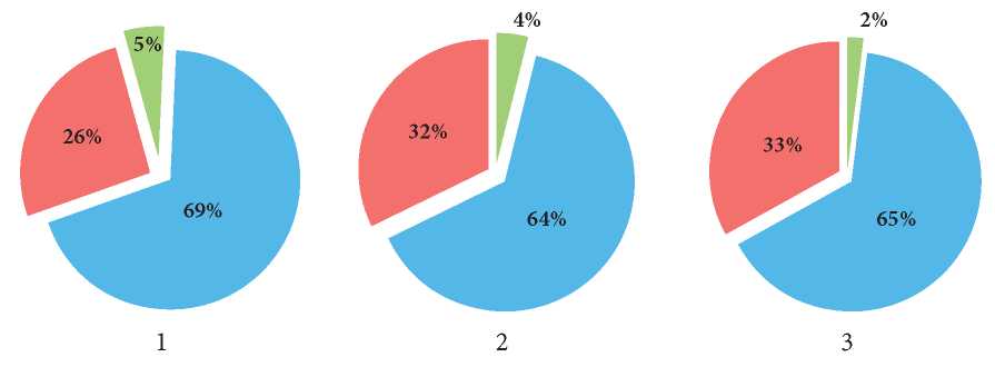
Fig. 2. Asymmetry of varying severity of the facial skull linear dimensions in women (%) within sigma 1 (blue), sigma 2 (red), and sigma (green) 3. 1 – upper “fan”, 2 – lower “fan”, 3 –lateral “fan”.
The authors revealed statistically significant differences in the degree of asymmetry in the facial skull linear dimensions in both sexes. In the upper “fan”, the distances from nasion to zygomaxilar (N-Zm) and from nasion to frontonazale (N-Fn) prevailed in both sexes on the right. In the lower “fan”, the distances from the supraspinal to zygomaxial (Ss-Zm) prevailed in women and men on the left side. In the lateral “fan”, the distance from the zygomaxial to frontonazale (Zm – Fn) prevailed on the right side in both sexes.

Fig. 3. SСT of the skull, front view (SSD image of shaded surfaces). The most asymmetric statistically significant linear dimensions of the facial skull in the examined men (1) and women (2) are shown by a black line in the upper “fan”, a white line – in the lower, and dashed lines – in the lateral “fan” on the left and right.
The revealed differences in the linear dimensions of the facial skull prevailed in men in comparison with women. These data corresponded to the data published by Alieva et al. [11].
SCT showed a prevalence of various asymmetries of the facial skull right-sided linear dimensions in men in all the “fans”. In women, right-sided asymmetry prevailed in the upper and lateral “fans” and left-sided asymmetry prevailed in the lower “fan”. These data do not agree with the respective data published by Alieva et al. [11] but confirm the data obtained by Gaivoronskiy et al. [13].
In the male and female groups, the sigma intervals of facial skull linear dimensions were used to evaluate the occurrence rate of asymmetry of varying severity. In the male and female groups, in the upper, lower, and lateral “fans”, there were differences revealed in parameters from both sides. Primarily, they did not exceed 1 or 2 ϭ. According to some authors [10][13], if the differences in the parameters from the left and right sides of the facial skull were within 1 ϭ, the revealed asymmetry was insignificant and physiological. If they were within 2 ϭ, the asymmetry was moderate, which did not require correction.
The analysis of SCT scans revealed sex-related differences in the linear dimensions of the facial skull. The majority of differences significantly prevailed in men.
SCT craniotomy in the upper, lower, and lateral “fans” revealed asymmetry of facial skull linear dimensions in men and women in the upper, lower, and lateral parts of the skull.
Right-sided asymmetry in the facial skull linear dimensions prevailed in all “fans” in men. In women, right-sided asymmetry prevailed in the upper and lateral “fans”, and left-sided asymmetry – in the lower “fan”.
The most expressed asymmetry in the facial skull linear dimensions was revealed in both sexes. In the upper “fan”, these dimensions were nasion-zygomaxial (N-Zm) and nasion-frontanazale (N-Fn) that prevailed in both sexes. In the lower “fan”, the asymmetry prevailed in the distance from supraspinal to zygomaxial (Ss-Zm) and from zygomaxial to frontanazale (Zm-Fn) in both sexes.
The revealed asymmetry in the facial skull linear dimensions of mild and moderate degree determines the individual shape of the face in both sexes. The obtained data on craniometry parameters can be used in clinical practice by radiologists, plastic surgeons, maxilla-facial surgeons, as well as anthropologists and forensic specialists.
1. Panina N.G., Perepelkin A.I., Krayushkin A.I. Modern views of facial asymmetry. Ural'skij medicinskij zhurnal. 2014;(7):126-129. (In Russ.) eLIBRARY ID: 22753142
2. Ko E.W.-C., Lin C.-H., Chen Y.-A, Chen Yu-R. Enhanced Surgical Outcomes in Patients with Skeletal Class III Facial Asymmetry by 3-Dimensional Surgical Simulation. Journal of Oral and Maxillofacial Surgery. 2018;76(5):1073-1083. DOI: 10.1016/j.joms.2017.09.009
3. Mareev O.V., Nikolenko V.N., Aleshkina O.U., Mareev G.O., Markeeva M.V., et al. Computer craniometry with the help of modern technology in medical craniology. Morphological newsletter. 2015;(1):49-54. (In Russ.) eLIBRARY ID: 25456984
4. Zhang D, Wang S, Li J, Zhou Y. Novel method of constructing a stable reference frame for 3-dimensional cephalometric analysis. Am J Orthod Dentofacial Orthop. 2018;154(3):397-404. DOI: 10.1016/j.ajodo.2017.11.038
5. Jiang X, Zhang Y, Bai S, Chang X, Wu L, Ding Y. Threedimensional analysis of craniofacial asymmetry and integrated, modular organization of human head. International Journal of Clinical and Experimental Medicine. 2017;10(8):11424-11431.
6. Park H., Lee J., Cho J., Hwang H., Lee K. Accuracy of three-dimensional cephalograms generated using a biplanar imaging system. Korean Journal of Ortthodontics. 2018;48(5):292-303. DOI: 10.4041/kjod.2018.48.5.292.
7. Dos Santos R.M.G., De Martino J.M., Haiter Neto F., Passeri A.L. Cone-beam computed tomography-based threedimensional McNamara cephalometric analyisis. Jornal of Craniofacial Surgery. 2018;29(4):895-899. DOI: 10.1097/SCS.0000000000004248.
8. Katsumuta A., Fujishita M., Maeda M., Ariji Y., Ariji E., Langlais R.R. 3D-CT evaluation of facial asymmetry. Oral Surg Oral Med Oral Pathol Oral Radiol Endod. 2005;99(2):212–220. DOI: 10.1016/j.tripleo.2004.06.072.
9. Kreutz M., Fitze B., Blecher C., Marcello A., Simon R., et al. Facial asymmetry correction with moulded helmet therapy in infants with deformational skull base plagiocephaly. Journal of Cranio-Maxillofacial Surgery. 2018;46(1):28-34. DOI: 10.1016/j.jcms.2017.10.013.
10. Gajvoronskij I. V., Dubovik E. I., Krajnik I.V. Morphometric parameters of facial cranium asymmetry in adult man. Morfologiya. 2009; 135(2);74-79 (in Russ.). DOI: 10.1016/j.jcms.2014.01.028.
11. Alieva SA, Shadlinsky VB, Movsumov NT. Sex-related features of the asymmetry of craniometrics parameters in various forms of the facial skull. Morfologicheskie Vedomosti – Morphological Newsletter. 2019;27(4):9-15. (In Russ.) DOI :10.20340/mvmn.19(27).04.9-15
12. Alekseev V.P., Debec G.F. Kraniometrija: Metodika antropometricheskih issledovanij. Moskva: Nauka.1964. 128 s.
13. Gajvoronskij I. V., Dubovik E. I., Krajnik I. V., Dergacheva E.A. Adult visceral cranium asymmetry and its assessment possibility. Vestnik Rossijskoj Voenno-medicinskoj akademii. 2009;1(25):140-144 (in Russ.). eLIBRARY ID: 12773593
14. Bahareva N.S. Features of the asymmetry of the linear dimensions of the facial skulls of residents of the South of Russia. Fundamental'nye issledovaniya. 2012;8-2:279-284. (In Russ.) eLIBRARY ID: 18304362
15. Efi mova У. Yu., Krayushkin A. I., Efi mov Yu. V., Bujanov E. A.. The linear parameters of the skull of mesocranial type. Volgograd Medical Scientifi c Journal, 2018;4(60):15-18. (In Russ.) eLIBRARY ID: 36784831
Elena V. Chaplygina, Dr. Sci. (Med.), Professor
Rostov-on-Don
Olga A. Kaplunova, Dr. Sci. (Med.), Professor
Rostov-on-Don
Maxim G. Shepetyuk, assistant
Rostov-on-Don
Olga P. Suhanova, assistant
Rostov-on-Don
Igor M. Blinov
Rostov-on-Don
Shepetyuk M.G., Kaplunova O.A., Shepetyuk M.G., Suhanova O.P., Blinov I.M. Sexual features of facial skull asymmetry according to spiral computed tomography. Medical Herald of the South of Russia. 2021;12(3):78-85. (In Russ.) https://doi.org/10.21886/2219-8075-2021-12-3-78-85
29, Nakhichevansky Lane, Rostov-on-Don, 344002
Rostov State Medical University
Тel.: +79185710558
e-mail: journal@medicalherald.ru


