


Contents
Scroll to:
https://doi.org/10.21886/2219-8075-2021-12-3-72-77
Scroll to:
Objective: To study the morphological features and subpopulation composition of immunocompetent cells of adhesion tissue in women with adhesions of the pelvic organs.
Materials and Methods: Th e study was carried out using surgical material obtained from 70 women aged 23 to 40 years. Of these, 50 tissue samples of peritoneal adhesions from patients with adhesions of organs in the small pelvis of I – II degree who underwent adhesiolysis and 20 samples of parietal peritoneum from healthy women who underwent endoscopic sterilization for contraception or completion of generative function. Th e authors used histological, immunohistochemical, and morphometric research methods.
Results: Immunological changes in adhesion tissue were characterized by the activation of the T-cell link of immunity. It was confi rmed by a signifi cant increase in the content of CD4+ (p <0.001), CD8+ (p <0.001), a shift in the balance of immunoregulatory subpopulations towards CD8+, a lower indicator of the immunoregulatory index (p = 0.015), and insuffi ciency of the humoral link of immunity, namely, the absence of CD20+ content against the background of a slight increase in the CD138+ pool.
Conclusion: To prevent the postoperative adhesion process in the small pelvis in patients of reproductive age, it is necessary to apply immunomodulatory therapy in the early postoperative period, which will improve the results of surgical treatment and is pathogenetically justifi ed.
Puchkina G.A., Sulima A.N., Davidova A.A. Immunocompetent cells in the tissue of adhesives of patients with adhesion process in the small pelvis. Medical Herald of the South of Russia. 2021;12(3):72-77. (In Russ.) https://doi.org/10.21886/2219-8075-2021-12-3-72-77
The adhesive process in the small pelvis is still an urgent issue of operative gynecology. It is proved that the adhesive process is a complication of up to 90% of all gynecological surgery cases [1][2]. Postoperative adhesions dramatically reduce the quality of patients’ life [3], as they lead to chronic pelvic pain [4] and infertility [5]. In recent years, while studying the pathogenesis of the adhesive process, some researchers have come to the conclusion that one of the links of adhesion formation is an altered immunobiological body reactivity [6][7]. However, to date, the role of the immune system in the pathogenesis of the adhesive process has not been fully studied.
The study of the cellular mechanisms of the formation of postoperative adhesions focused mainly on the role of peritoneal macrophages, polymorphonuclear neutrophils, and cytokines [7][8][9]. In previous studies, the leading role of T-lymphocytes in the coordination and regulation of chemotactic responses in various infectious, autoimmune and inflammatory tissue diseases has been proved [10][11][12]. However, there are isolated experimental studies [13][14], which provide data on the importance of T-lymphocytes in the development of the postoperative adhesive process, which are insufficient for unambiguous conclusions.
The immune mechanisms of the pathogenesis of adhesion formation are based on violations of the recruitment processes of monocytes that differentiate into macrophages and secrete a number of cytokines, chemokines, and growth factors, with the involvement of humoral immunity cells – plasmocytes and their precursors B-lymphocytes. This causes pathological tissue regeneration in the focus of inflammation [9]. At the same time, the authors of this article have not found any works in the available literature devoted to the study of the role of the subpopulation composition of B-lymphocytes in the development of postoperative adhesions.
Thus, according to the data on the immune system importance in the regulation of the inflammation explicitness and its outcome, the authors of this study assumed that changes in the composition and amount of expression of immunocompetent cells are crucial in the inflammatory process formation. They lead to the development of the postoperative adhesive process of the pelvic organs.
The aim of the study was to study the morphological features and subpopulation composition of immunocompetent cells of the adhesions tissue in women with adhesions in the pelvis. The study was approved by the Ethical Committee of the S. I. Georgievsky Medical Academy of the V. I. Vernadsky Crimean Federal University in accordance with Order No. 12/4/107 dated June 16, 2021, planned and conducted in accordance with the Helsinki Declaration. All the patients with the adhesive process in the small pelvis and healthy women included in the study signed a voluntary informed consent.
The study was performed on surgical material obtained from 70 women aged 23 to 40 years (their average age was 36.2 [ 33.1–37.2]). Therefore, fifty (50) tissue samples of peritoneal adhesions were taken from patients with adhesions of organs in the small pelvis (stages I–II stage) who underwent adhesiolysis, and twenty (20) samples of parietal peritoneum were taken from healthy women who underwent endoscopic sterilization for the purpose of contraception or completion of generative function.
Histological examination of the adhesions tissue and peritoneum was performed according to the standard method. Hematoxylin and eosin staining was also used for a review assessment of the structural tissue features.
Immunohistochemical (IHC) tissue study was performed according to a standardized method – prepared micro-preparations of tissue samples taken from adhesions and peritoneum were used. The CD4+, CD8+, CD20+, CD138+ lymphocytes identification was performed while using a panel of four monoclonal mouse antibodies (Clone 4B12 Ready-to-Use, CloneC8/144B Ready-to-Use, Clone L26 Ready-to-Use, Clone MI15 Ready-to-Use (DAKO company)) and NovocastraNovolinkTM imaging systems based on the compact polymer NovolinkCompactPolymer™ (Leica, Germany) on the BondMax IHC stainer (Leica, Germany). In each section, the absolute number of immunocompetent cells labeled with the corresponding antibodies was calculated in 10 randomly selected fields of vision at an increase of 400 using the Software DP-SOFT program. Two (2) sections on 1 mm were listed.
The micro-preparations were viewed and photographed using an Olympus 5050Z digital camera mounted on an Olympus CX-41 microscope.
Statistical processing of the obtained data was performed by means of the STATISTICA 8.0 application software package. The average value (M) and standard deviation (σ (±)) were used as descriptive statistics for quantitative values. The inter-group comparison was carried out using Student's t-test. The differences were considered statistically significant at p < 0.05.
While studying the adhesive tissue obtained during surgery, it was found that pelvic peritoneal adhesions were arrays of connective tissue with weak signs of interstitial edema, partially covered with mesothelium. The consistency of the adhesions was loose and fibrous with focal fibrinoid changes and hyalinosis. The integumentary mesothelium consisted of flattened cells of a cubic shape or formed a kind of “pillows” represented by proliferating mesotheliocytes. As part of the adhesions, thin nerve stems, focal weakly or moderately pronounced, mainly perivascular mononuclear infiltration were visualized. In some cases, adipocytes forming focal clusters or completely sprouting a spike were also included in the composition of the adhesion tissue. In the thickness of the adhesions, bundle growths of vessels of the microcircular bed were also detected, which are mainly arterioles with a violation of the architectonics of endotheliocytes, which was manifested in their perpendicular arrangement relative to the vessels intima. Blood capillaries were, as a rule, full-blooded; in some cases, the phenomena of stasis and erythrocyte sludge were observed in them.
During the IHC study of the adhesive tissue, it was found out that CD4+ T-lymphocytes were located in the form of band-shaped infiltrates and focal perivascular clusters (Figure 1A). CD8+T-lymphocytes were located mainly submesothelially, in the form of perivascular clusters (Figure 1B).
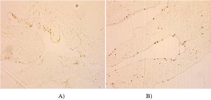
Figure 1. Fragment of peritoneal adhesions.
Notes: A – stripe infiltrates from CD4 + T-lymphocytes. IHC. Staining with hematoxylin and eosin. Uv. 100×. B – submesothelial CD8 + T-lymphocytes. IHC. Staining with hematoxylin and eosin. Uv. 100×.
During the IHC study of the adhesive material, it was found that CD138+ were located mainly in a perivascular way, as part of mononuclear infiltrates (Fig. 2).
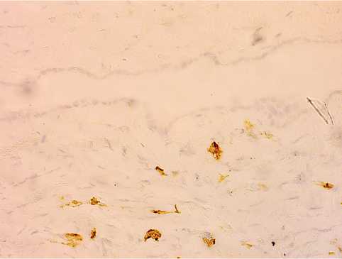
Figure 2. Fragment of peritoneal adhesion with focal accumulations of CD138 + cells in adhesion tissue. IHC. Staining with hematoxylin and eosin. Uv. 100×.
It should be noted that CD20+B-lymphocytes were not detected in the tissue of adhesions.
The morphometric analysis of the subpopulation composition of immunocompetent tissue cells of adhesions in women with adhesions in the pelvis and peritoneum in healthy women obtained the data presented in Table 1.
Table 1
Comparative analysis of the expression of immunocompetent cells in adhesion tissue and peritoneum
|
Indicators |
Adhesion tissue, units of inc. |
Peritoneum, units of inc. |
p |
|
CD4+ |
5.6±0.5 |
3.1±0.2 |
P<0.001 |
|
CD8+ |
9.2±0.6 |
3.0±0.3 |
P<0.001 |
|
CD4+/ CD8+ |
0.6±0.02 |
1.1±0.2 |
P=0.015 |
|
CD20+ |
0 |
2.1±0.1 |
P<0.001 |
|
CD138+ |
0.8±0.2 |
0.6±0.1 |
P=0.580 |
In the adhesions tissue, compared with the peritoneum, a higher statistically significant number of CD4+T-lymphocytes, CD8+T-lymphocytes, and the absence of CD20+-B-lymphocytes were visualized. The number of CD138+ cells was slightly increased, but did not reach statistical significance.
The immune system plays a major role in regulating the explicitness of inflammation and its outcome [15]. It is known that it is the functional state of the cellular and humoral immune system components that largely determines the course and outcome of the inflammatory process in the abdominal cavity [16].
The present study is devoted to the features of the phenotypic composition of immunocompetent cells in the adhesion tissue in women with the adhesive process in the pelvis (stages I–II).
The peritoneal immune compartment is a microenvironment with a certain repertoire of various subpopulations of lymphocytes that trigger a cascade of molecular processes [17]. According to modern concepts, the leading place in the initiation of an immunological reaction belongs to the activation of a pool of antigen-recognizing CD4+T-helper cells that determine the nature and intensity of the immune response [18]. As the results of this study showed, a higher statistically significant number of CD4+T-lymphocytes is registered in the tissue of the adhesions compared to the peritoneum. The data obtained by the authors of this article are close to the results of the experimental work of Doo Ryeon Chung et al. [13], which shows that CD4+T-lymphocytes play a significant role in the development of the adhesive process. Pro-inflammatory cytokine IL-17, produced mainly by activated CD4+T-helper cells, inducing the release of CXC-chemokines, macrophages-inflammatory protein-2, cytokine-induced neutrophils chemoattractant, indirectly affects the development of postoperative adhesion. The authors also found that the severity of the adhesive process directly depended on the quantitative composition of the pool of CD-4+T-lymphocytes. The obtained data indicates the significant role of immunocompetent cells in the formation of postoperative adhesions.
Therefore, CD-8+T-lymphocytes are the main effector unit of cell-mediated immunity and play an essential role in the formation of natural or antigen-induced (surgical intervention) immunological tolerance. While performing lysis, CD-8+T-lymphocytes ensure the genetic constancy of the internal environment of the body. In the present study, a higher statistically significant number of CD8+T-lymphocytes in the adhesions tissue compared to the peritoneum was recorded. This fact has not been discovered by the authors of this article in any similar work, so it needs additional study. However, based on modern ideas about the mechanisms of the immune response in the postoperative period, it can be assumed that a violation of the function of the T-suppressor immune system component suppresses the recruitment of other immunologically active cells (mainly monocytes and neutrophils) into the focus of vascular bed restructuring. In turn, the subsequent violation of the production of mediator molecules, peptide growth and anti-growth factors, the expression of leukocyte and platelet adhesive molecules serves as an additional factor of pathological adhesion.
The immunoregulation index (CD4+:CD8+ ratio) is an indicator of the harmonious functioning of the immune system and is normally 1:2. In the present study, in patients with the adhesive process, the ratio of CD4:CD-8 in the adhesions tissue was 0.6, which indicates the insufficiency of the local immune response. The shift of the ratio to the left occurred due to a decrease in the number of T-helpers and an increase in the number of T-suppressors.
CD-20+B-lymphocytes with regulatory functions are found in tissues in a number of inflammatory and autoimmune diseases, where their role is mainly reduced to the suppression of pathological processes. Normally, SD20+ B-lymphocytes differentiate into plasma cells, as they synthesize immunoglobulins, the final stage of which is the binding and leveling of the antigen. In this study, the absence of CD20+ B-lymphocytes in the tissue of adhesions was established, which indicates the lack of functional activity of the humoral link of immunity. This fact has not been discovered in any similar work and needs additional study as well.
CD-138+ (syndecan-1) is a universal marker of plasma cells. It was found that CD138+ was directly involved in the development of intercellular connections and blocks the penetration of cells into collagen. At the same time, given the role of CD138+in the attachment of epidermal growth factors to cells, the absence of this molecule on the cell membrane may reduce their susceptibility to the bioregulatory action of authentic cytokines. In the present study, a higher amount of CD138+ was recorded in the adhesions tissue compared to the parietal peritoneum, but it did not reach statistical significance. The obtained result, in the authors’ opinion, indicates a continuing slow regeneration of the tissue with a transient manifestation of IHC markers of inflammation.
Thus, a significant imbalance of immunoregulatory processes was revealed in patients with the adhesive process of the pelvic organs at the local level. It was characterized by a violation of the functional activity of the cellular and humoral immune system components. It is proved that the predominant influence on the process of adhesion is exerted by the T-cell immune system components. In order to prevent the postoperative adhesive process in the pelvis in patients of reproductive age, it is necessary to use immunomodulatory therapy (sodium deoxyribonucleate) during the early postoperative period, which is pathogenetically justified. This will improve the results of surgical treatment.
1. Shatova E.S. Modern approach to the problem of adhesive disease in women of reproductive age. J. obs. and fem. dis. 2013;62(1):90-101. (In Russ.) eLIBRARY ID: 19409693
2. Dobrokhotova Y.E. Grishin I.I. Grishin A.I. Experience of using an anti-adhesion barrier in patients with tuboperitoneal factor infertility. RMJ. 2017;25(15):1141–1143. (In Russ.) eLIBRARY ID: 30309182
3. Bezhenar' V.F., Aĭlamazian É.K., Baĭliuk E.N., Tsypurdeeva A.A., Polenov N.I. Etiology, pathogenesis and prevention of adhesions in surgery of the pelvic. Russian Obstetrician-Gynecologist Gazette. 2011;11(2):90-101. (In Russ.) eLIBRARY ID: 18965970
4. Bezhenar V.F., Tsypurdeeva A.A., Baylyuk E.N. Adhesive disease pelvic organs in gynecological patients: from pathogenesis to prevention. Oncogynecology. 2014;(4):68–74. (In Russ.) eLIBRARY ID: 22752601
5. Burlev V.A., Dubinskaia E.D. Phenotypic features of undiff erentiated forms of connective tissue dysplasia in patients with pelvic peritoneal adhesions. Russian journal of human reproduction. 2012;18(2):8-14. (In Russ.) eLIBRARY ID: 18022550
6. Lazarenko V.A., Konoplia A.I., Lipatov V.A., Gomon M.S., Efremenkov A.M. To the question of role of the immune system in the development of the adhesion process of abdominal cavity (literature review). Innova. 2016;5(4):29–33. (In Russ.) eLIBRARY ID: 35550936
7. Rybalka A.N., Sulima A.N., Davydova A.A., Yakovchuk Y.K., Anikin S.S. Features of proinfl ammatory cytokines expression in pelvic adhesions tissue at women with pelvic chronic infl ammatory diseases. Juvenis Scientia. 2016;(3):29-31. (In Russ.) DOI 10.15643/jscientia.2016.3.101.
8. Kuraoka S, Campeau JD, Nakamura RM, diZerega GS. Modulation of postsurgical macrophage function by early postsurgical polymorphonuclear leukocytes. J Surg Res. 1992;53(3):245-50. doi: 10.1016/0022-4804(92)90042-x
9. Magomedov M.A. Local cellular regulation in the formation of postoperative adhesions in peritonitis. Pirogov Russian journal of surgery. 2004;(6):9-11. (In Russ.) eLIBRARY ID: 35540526
10. Torgashina A.V., Biykovskaya S.Y., Soloviev S.K., Nasonov E.L. T-regulatory cells in systemic lupus erythematosus and rheumatoid arthritis. Rheumatology Science and Practice. 2009;47(3):50-59. (In Russ.). eLIBRARY ID: 20342153
11. Diani M, Altomare G, Reali E. T cell responses in psoriasis and psoriatic arthritis. Autoimmun Rev. 2015;14(4):286-92. DOI: 10.1016/j.autrev.2014.11.012
12. Griveas I, Fleva A, Karanikas E, Gogos K, Sakellariou G. CD4/CD8 T-cell ratio in peritoneal dialysis effl uents predicts the outcome of peritonitis in patients undergoing continuous ambulatory peritoneal dialysis. Artif Organs. 2009;33(12):1091-5. DOI: 10.1111/j.1525-1594.2009.00802.x
13. Chung DR, Chitnis T, Panzo RJ, Kasper DL, Sayegh MH, Tzianabos AO. CD4+ T cells regulate surgical and postinfectious adhesion formation. J Exp Med. 2002;195(11):1471-8. DOI: 10.1084/jem.20020028
14. Dong L, Zheng X, Wang G. Peritoneal adhesions induce Th 17/Treg imbalance in mice. Int J Clin Exp Pathol. 2018;11(9):4352-4362. PMID: 31949832; PMCID: PMC6962961.
15. Lebedeva O.P., Pakhomov S.P., Karpov P.A. Churnosov M.I., Popov V.N. Signifi cance of the innate immunity Toll-like receptors in development of obstetric and gynecological disorders. Immunology, allergology, infectology. 2012;(1):19–26. (In Russ.). eLIBRARY ID: 21165785
16. Lazarenko V.A., Lipatov V.A., Gomon M.S., Efremenkov A.M. To the question of role of the immune system in the development of the adhesion process of abdominal cavity (literature review). Innova. 2016;4(5):29-33. (In Russ.). eLIBRARY ID: 35550936
17. Kulakov V.I., Volkov N.I., Bespalov G.V. Immunocompetent cells in the endometrium of patients with infertility and external genital endometriosis. Journal of obstetrics and womans diseases. 2003;52(4):12-16. (In Russ.). eLIBRARY ID: 9231996
18. Babayeva A.G. Regeneration and immunogenesis system. Moscow: Medicine; 1985. (In Russ.).
Galina A. Puchkina, Assistant, Department of Obstetrics, Gynecology and Perinatology № 1
Simferopol, Republic of Crimea
Anna N. Sulima, Dr. Sci. (Med.), Professor, Department of Obstetrics, Gynecology and Perinatology № 1
Simferopol, Republic of Crimea
Aleksandra A. Davidova, Cand. Sci. (Med.), Assistant Professor of department of pathlogical anatomy with section course
Simferopol, Republic of Crimea
Puchkina G.A., Sulima A.N., Davidova A.A. Immunocompetent cells in the tissue of adhesives of patients with adhesion process in the small pelvis. Medical Herald of the South of Russia. 2021;12(3):72-77. (In Russ.) https://doi.org/10.21886/2219-8075-2021-12-3-72-77
29, Nakhichevansky Lane, Rostov-on-Don, 344002
Rostov State Medical University
Тel.: +79185710558
e-mail: journal@medicalherald.ru


